INTRODUCTION
Methods for repairing skeletal deficiencies now include cell-based technologies.
Current research efforts aim to develop new approaches for generating
tissues in vitro that would integrate in vivo focus on cell carriers or
scaffolds and methods to maintain the phenotype of the cells and to promote
3D histogenesis. Towards these ends, we developed porous 3D collagen sponge
scaffoldings to deliver different cells and to support histogenesis. In
addition, we used a simple system of medium perfusion and a more complex
computer-driven system for application of fluid pressure for histogenesis
in vitro.
 |
|
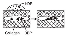
| Figure 1. Representation of in vitro chondroinduction. Human dermal fibroblasts (hDF) are seeded on top of the collagen sponge which contains a filling of demineralized bone powder (DBP, 75-250 µm) . The hDFs migrate through the collagen layer and those that invade the DBP are induced to produce cartilage matrix. |
THREE-DIMENSIONAL
CULTURE OF CELLS
Monolayer, or two-dimensional, culture of disaggregated, anchorage-dependent
cells in vitro has allowed for progress in understanding regulation of
cell differentiation, growth, and function as well as cell-to-cell interactions
that modulate these processes. Because monolayer culture does not model
for the architecture of tissues and organs, analysis of integrated function
or dysfunction of tissues or organs requires a different mode of experimentation.
One approach is to retain cellular distributions within a block of tissue
and to maintain it intact in what is called "organ culture."
The major challenge in organ culture is to assure sufficient gas and nutrient
exchange in order to maintain viability of cells throughout the tissue
mass. A second approach is to grow disaggregated cells to high density
with a three-dimensional (3D) geometry. Organoid structures will grow
if kept supplied with fresh medium either by gyration or perfusion. Use
of scaffolds can enhance histogenesis. Various materials have been tested
as scaffolds to support the growth of musculoskeletal cells, including
collagen, hydrogels, resorbable synthetic polymers, and calcium phosphates.
Our laboratory uses collagen fibers prepared as a porous lattice;these
have superior biocompatibility, reproducibility, and low cost. We reported
that some preparations of collagen should be avoided because of their
incompatibility in vivo. 1
3D
CULTURE OF CHONDROCYTES
Cartilage has been a model for research of in vitro engineered tissues
on scaffolds because of its cellular homogeneity and avascularity. 2
On the other hand, chondrocytes require certain conditions to ensure that
they maintain the features of differentiated, matrix producing cells when
cultured in vitro . 3
Histochemical, immunohistochemical, and immunochemical analyses showed
that cartilage matrix can accumulate in porous collagen sponges seeded
with bovine articular chondrocytes. 4
Analysis of the cartilage-specific genes, aggrecan and collagen type II,
have demonstrated preservation of the chondrocyte molecular signature
compared to dedifferentiation in monolayer culture. 5
Preliminary investigations indicated that articular and endochondral chondrocytes
retain their differences in these collagen sponges. 6
The collagen sponge
has been used to determine the effects of insoluble, soluble, and mechanical
factors on chondrogenesis. For example, hyaluronan, another important
constituent of cartilage matrix, was incorporated into the porous collagen
sponges. 7
A small amount of hyaluronan (2% w/w) in 3D collagen scaffolds enhanced
chondrogenesis, but a greater amount was found to be inhibitory. This
finding lends support to the idea that biomimetic, or "smart"
scaffolds can be developed with optimal signals for specific histogenesis.
CHONDROINDUCTION
IN VITRO
Demineralized bone has been used clinically and in experimental animals
to induce endochondral osteogenesis. 8
The first steps appear to be migration of nearby connective tissue cells
to the demineralized bone and their transdifferentiation into chondrocytes.
In an attempt to identify the mechanisms of chondroinduction, we cultured
human dermal fibroblasts in collagen sponges that contained particles
of demineralized bone (Figure 1) . One
of the most critical parameters for chondroinduction is a high packing
density of the demineralized bone. 1,
9 Histochemical, immunohistochemical, and immunochemical evidence
documented that after culture with demineralized bone, human dermal fibroblasts
produced cartilage extracellular matrix (ECM) (4) and expressed cartilage-specific
matrix genes. 8
Analysis of gene shifts prior to full expression of the cartilage matrix
genes should reveal controlling genes that are specifically up-regulated
by exposure to demineralized bone. Recently, a new method of comparing
cellular expression of genes, Representational Difference Analysis (RDA)
, was used to distinguish a pool of genes that were up-regulated during
the initial steps of chondroinduction of human fibroblasts. 10
The identified genes represented several functional classes, including
cytoskeletal elements, protein synthesis and trafficking molecules, and
transcriptional regulators. Some of the identified genes are novel and
others are known sequences with unknown functions. Thus, the culture system
has the power to reveal important changes that forerun chondroblastogenesis.
3D CULTURE OF HUMAN MARROW CELLS
Bone marrow contains adherent cells that give rise to osteoblasts and
non-adherent cells that give rise to osteoclasts. Marrow was obtained
during the course of total hip arthroplasty from 39 men aged 37-86 years.
11 Cells
were cultured in porous collagen sponges and were assessed for alkaline
phosphatase activity, as a marker of osteoblast differentiation. A quantitative,
competitive reverse transcription-polymerase chain reaction (RT-PCR) assay
showed that patients <50 years of age had 3-fold more mRNA for
alkaline phosphatase than those >60 years of age (p =0. 021)
. Pearson correlation indicated a significant decrease in mRNA for alkaline
phosphatase with age (r =-0. 78, p =0. 028) . These molecular and histoenzymatic
data suggest that the osteogenic potential of human bone marrow cells
decreases with age.
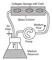 |
Figure 2. Schematic of the system to perfuse medium through the porous collagen sponges. Sponges are suspended in medium in a glass chromatography column. A peristaltic pump perfuses medium through the system. All components except the pump are held in a 37 ¡C incubator. |
|
Different culture conditions are needed for the differentiation of bone-resorbing osteoclasts. Recently, it was shown that osteoclastogenesis is inhibited by a product of osteoblasts or marrow stromal cells, called osteoprotegerin (OPG) . We tested whether age influenced OPG expression using bone marrow cells from 18 subjects, aged 38-84 years. 12 Expression of OPG in the younger group was 500% greater than in the older group (p = 0. 034) . Decline in the expression of OPG with age may increase the capacity of stromal/osteoblast cells to support osteoclastogenesis.
Human marrow stromal
cells also have the capacity to differentiate into chondrocytes. Molecular
analysis of chondrocyte and adipocyte genes expressed in marrow cells
cultured in 3-D collagen sponges showed that TGF-B1 promoted chondrogenesis
and inhibited adipogenesis. 13
EFFECTS
OF MEDIUM PERFUSION ON HISTOGENESIS
Once cells proliferate within 3D scaffolds, dense aggregates of cells
and the accumulation of ECM may impede transfer of nutrients and wastes.
Recently, we reported that medium perfusion enhanced the viability and
function of murine bone marrow cells 14
with a perfusion culture system made from standard laboratory equipment
(Figure 2) . One advantage of 3D collagen
sponge scaffolding is the ease of combining multiple cell types. Murine
marrow stromal cell and IL-3-dependent hematopoietic cells were co-cultivated
within collagen sponges. Viability of these cells was poor under standard
conditions. Perfusion of medium, however, stimulated both the proliferation
of the stromal cells and their ability to support the growth of the factor-dependent
hematopoietic cells.
The perfusion system enhanced histogenesis by bone-forming cells, murine K8 osteosarcoma cells, maintained in 3D collagen sponges. 15 With perfusion, there was greater viability, more alkaline phosphatase-positive cells, and more mineralized tissue after 21 days than in non-perfused control sponges. Quantitative measures of cell proliferation, DNA content, calcium accumulation, and expression of the bone-specific genes, collagen type I and osteocalcin, were dramatically increased with perfusion. Obstacles to clinical application of engineered bone include optimization of starting cells, tissue vascularization, and both acute and long-term compatibility of scaffolds or their degradation products. 16
Recently, we demonstrated that perfusion profoundly increased the viability and growth of human oral mucosal cells on porous scaffolds. 17 Clinically, thin layers of engineered epithelial tissue are technically difficult to transplant. With perfusion for one week, the keratinocytes formed multiple layers almost twice as thick as without perfusion.
Efficient medium exchange is not beneficial to all cell types. Because articular cartilage is avascular and exposed to relatively poor nutrient and low oxygen conditions, we wondered whether chondrocytes in 3D scaffolds would benefit by medium perfusion. Bovine articular chondrocytes (bACs) were grown in 3D collagen sponges with or without medium perfusion (0. 33 ml/min) for up to 15 days. 18 The influence of medium perfusion was evaluated using markers of cartilage matrix accumulation, synthesis, and gene expression. These measures showed significantly better chondrogenesis without perfusion. Thus, the perfused conditions that are beneficial for other cell types inhibit chondrogenesis.
Because increased oxygen exchange may have been deleterious to the chondrocytes, we tested low oxygen concentration (tension) in this system. 19 In the growth plate, oxygen tension is low in the reserve zone (20 mm Hg;2. 6% ) and highest in the proliferative zone (57 mm Hg;7. 6% ) . Even during fracture repair, oxygen tension varies considerably (~20-30 mm Hg;2. 6 -4% ) during callus formation. Collagen sponges were exposed to medium perfusion at either 2% or 19% (atmospheric) oxygen concentrations. Matrix synthesis was greater without perfusion in standard conditions of 19% oxygen. Reduction to 2% resulted in 130% increase in matrix synthesis. Thus, chondrogenesis was restored by reduction of oxygen concentration in the perfused medium to 2%.
With this system,
the shear stress caused by medium perfusion at 1. 3 ml/min was 0. 00157
dynes/cm 2 , which is 0. 1-1% of the magnitude of shear stress achieved
in veins. In the experiments with flow rate at 0. 3 ml/min, the shear
stress was 0. 00037 dynes/cm 2 . Even at these low shear stresses, cell
surface receptors such as integrin could theoretically be stimulated,
with subsequent signal transduction to regulate gene expression. The mechanisms
of such mechanotranduction are the subject of intense research. Our
homemade culture system is capable of defining these dynamic mechanisms.
EFFECTS
OF FLUID PRESSURE ON CHONDROGENESIS IN POROUS COLLAGEN SPONGES
All skeletal tissues are under compressive loading and stretching tension.
We designed a novel pressure/perfusion culture system to apply fluid pressure
(FP) to the medium perfusing the sponges. 20 The magnitude of pressure
was 2. 8 MPa, which was within the physiological range of 0 -3. 5 MPa
that is achieved at the knee during normal walking. After 15 days, there
was 300% more accumulated matrix with FP, applied either continuously
or intermittently (0. 015 Hz) . Matrix synthesis was 40% greater with
FP than control. With this novel fluid pressure culture system, 2. 8 MPa
fluid pressure stimulated synthesis of cartilage specific proteoglycans
in chondrocytes cultured in 3D collagen sponges.
These 3 platform technologies -porous collagen sponges, the perfusion system, and fluid pressure apparatus are being used to optimize 3D histogenesis of different tissues. Rational manipulation of soluble factors, insoluble factors, and physical factors are expected to result in greater understanding of the cellular and tissue mechanisms of growth and in practical applications for repair of skeletal tissues.
Julie Glowacki, PhD is an Associate Professor in Orthopaedic Surgery at Harvard Medical School.
Shuichi Mizuno, PhD, Research Staff, Brigham and Women's Hospital Orthopaedics Research Laboratories, Boston.
Address correspondence to:
Julie Glowacki, PhD
Orthopaedic Research Laboratory
Brigham and Women's Hospital
75 Francis Street
Boston, MA 02115