Dr. Mehmet zbaydar, a visiting fellow from Turkey, spent six months with us and participated in a study that evaluated various repair techniques for the rotator cuff tears. He has returned to his clinical practice in Istanbul, Turkey. Dr. Markus Tingart, an orthopedic resident from Cologne, Germany, is doing a research fellowship for 15 months and studies the bone quality and architecture of the proximal humerus and their effect on strength of the rotator cuff reconstructions. Markus received a prestigious research fellowship grant from DFG, German Research Society, (equivalent of the NIH) to do this project. He is also involved in the study that evaluates the volume of the rotator cuff muscles using MRI. He started this project together with Dr. Alex Poon, an orthopedic resident from Hong Kong and is now working with Dr. Jenne Lehtinen, a visiting fellow from Finland.
Dr. Ariane Gerber is from Switzerland and has been selected to become the first Harvard-Zurich shoulder fellow. After spending 6 months in Zurich, Switzerland, she continued her shoulder fellowship with Dr. Warner and subsequently worked in the Hand and Upper Extremity Service with Dr. Jesse Jupiter. She now has a temporary faculty position with the Harvard Shoulder Service. Despite her strong involvement in the clinical care of patients, she found time to perform important research studies in the area of complex reconstructive procedures of the shoulder joint. Dr. Gerber received a research grant from the Swiss Science Foundation for one of the projects that investigates various tendon transfer procedures around the shoulder.
It has also been a pleasure to work with two medical students, Peter Fitzgibbons, who is now at Dartmouth Medical School, and Fraser Harrold from Dundee University School of Medicine in Scotland. With their engineering and science background, both have provided significant contributions to several of our projects. Fraser has maintained his interest in orthopedic science and is planning to return to Boston for the third time this summer to continue working in the area of shoulder biomechanics.
Rotator
Cuff Tears:The Effect of Reconstruction Method on Three-Dimensional Repair
Site Area
M
Apreleva, M zbaydar, P Fitzgibbons, JJP Warner
Structural failure
of the rotator cuff repair is the most frequently encountered complication
with the majority of reruptures occurring at the tendon-bone interface.
Healing and strength of the repair may depend on the size of the attachment
area between tendon and bone after the repair. However, little is known
about the repair site areas after surgical reconstructions and controversy
still exists as to whether suture anchor repair results in stronger tendon
to bone fixation than transosseous repair. We quantitatively compared
and visualized the threedimensional area of the original supraspinatus
insertion with the repair site area after four reconstructions of a simulated
supraspinatus tear.
 |
|
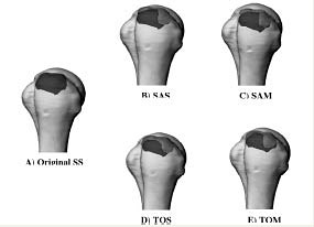
| Figure 1. 3D insertion site and repair site areas of the SS tendon superimposed onto a computer model of the humeral head. A) Original SS insertion. Repair site areas superimposed onto the original SS insertion:B) Suture anchor mattress repair, C) Suture anchor simple repair, D) Transosseous mattress suture repair, E) Transosseous simple suture repair. |
The outline of the original supraspinatus (SS) insertion was obtained in 10 human cadaveric shoulders using a 3D digitizer. A supraspinatus tear was created and four repair techniques were evaluated:transosseous simple suture (TOS) , transosseous mattress suture (TOM) , suture anchor simple suture (SAS) , suture anchor mattress suture (SAM) . The 3D outlines of the reconstructed supraspinatus insertion were digitized after each repair. The outlines of the original supraspinatus insertion and repair areas were superimposed onto humeral geometry obtained from a laser scanner and surface areas were calculated.
We found that the original supraspinatus insertion area was larger than any of the repair site areas (Figure 1) . On average, transosseous simple suture repair provided 20%larger repair site area than the other repairs. Repair site areas were not different among transosseous mattress, suture anchor simple and suture anchor mattress repairs, and covered 67%of the original SS insertion. Our results suggest that transosseous simple suture fixation may provide greater potential for osseous incorporation and healing at the tendon-bone interface by increasing the repair site area, and thus, greater strength of the repair. With the development of new biological enhancement techniques, it might prove important to maintain a large area of contact between tendon and bone, allowing more fibers to participate in healing process. This study also helps to better understand the three-dimensional rotator cuff insertion anatomy and may improve treatment and prevent failures of rotator cuff surgeries.
While many factors influence the outcomes of the rotator cuff repairs and optimum surgical technique is yet to be found, based on this study we conclude that attention should be paid to the three-dimensional geometry of the repair and the ability of the procedure to restore original tendon insertion providing improved strength by creating increased tendon-to-bone attachment area.
Characterization
of the Three-Dimensional Bone Mineral Density Distribution in the Proximal
Humerus
M
Tingart, M Apreleva, JJP Warner
Twenty-five
million Americans currently suffer from osteoporosis and 80%of those afflicted
are women. It has been shown that decreased bone quality may result in
increased risk for fractures and poor fixation of the implants, regardless
of the surgical technique chosen to treat these fractures. Approximately
300, 000 proximal humeral fractures occur annually in the US alone and
60, 000 require surgical treatment. Postoperative implant loosening, fracture
re-displacement and impaired fracture healing are complications commonly
seen in patients with osteoporosis. Poor bone quality also affects the
surgical treatment of rotator cuff tears in the shoulder. In osteoporotic
bone, transosseous sutures may cut through the bone or suture anchors
may pull out of the bone before tendon healing, and result in failure
of the repair.
We investigated the three-dimensional distribution of total, trabecular and cortical bone mineral density (BMD) in the head and the surgical neck of the humerus using Dual Energy X -Ray Absorptiometry and peripheral Quantitative Computer Tomography. We found an overall decrease in total, trabecular and cortical BMD from the proximal to the distal part of the humeral head. Furthermore, there are significant differences in BMD between the anterior and posterior part of the articular surface as well as between the greater and lesser tuberosity. In addition, there are regions of significantly different cortical and trabecular BMD within the area of the greater and lesser tuberosity.
Besides bone density, trabecular microarchitecture of the humeral head might be important to detect differences in the biomechanical properties of various parts of the humeral head. Currently, we are analyzing the bone microarchitecture of the humeral head by Micro-CT with a resolution of 60 µm. After this quantitative assessment of BMD and trabecular microarchitecture, we are planning to determine the effect of these bone quality parameters on the strength of fixation of the rotator cuff suture anchors and internal fixation devices used for proximal humeral fractures. This information will help to understand osteoporosis related fractures and to improve current treatment options for these injuries.
Reliability
and Validity of the Magnetic Resonance Imaging in the Quantitative Assessment
of Rotator Cuff Muscle Volume
M
Tingart, M Apreleva, J Lehtinen, JJP Warner
Chronic progression
of rotator cuff tears is accompanied by muscle atrophy, fatty degeneration
and retraction. These tendon and muscle changes often result in re-rupture
of the repair and adversely affect the functional outcome after rotator
cuff surgery. It has been suggested that changes in muscle volume are
related to muscle quality and function. Therefore, preoperative quantitative
assessment of rotator cuff muscle volume is clinically relevant for surgical
planning and prediction of outcome after rotator cuff repair. However,
little information is available on the reliable and valid method to estimate
muscle volumes in-situ. The objective of this study was to evaluate the
reliability and validity of the Magnetic Resonance Imaging (MRI) for quantitative
determination of rotator cuff muscle volume.
Muscle volumes of the rotator cuff muscles were determined in 10 human cadaveric shoulders using two methods. First, sagittal MRI scans of each shoulder were obtained and three investigators independently traced the contours of the supraspinatus, infraspinatus/teres minor and subscapularis muscles three times. Muscle volumes were calculated using special 3D image analysis software. Rotator cuff muscles were then dissected and volume of each muscle was measured by water displacement. The muscle volume measured by two methods was compared using Pearson correlation. Intra-and inter-observer reproducibility and reliability were also determined. Our preliminary results indicate a good correlation between the muscle volume measured using the MRI technique and the volume measured with the water displacement method.
The
Value of Pectoralis Major Tendon as an Anatomical Landmark to Determine
Humeral Length and Retroversion
A Gerber, M Apreleva, F Harrold, JJP Warner
When hemiarthroplasty of the shoulder is required to reconstruct comminuted
fractures or malunions, proper positioning of the humeral component is
difficult. The pectoralis major tendon is a well-defined structure that
is visible during a delto-pectoral approach. Therefore, we hypothesize
that this tendon can be used as a reference point to determine the proximal
humeral anatomy when performing an arthroplasty procedure.
 |
|
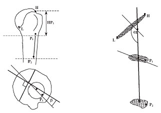
| Figure 2. A) Schematic of the anterior and superior views of the proximal humerus with digitized and calculated anatomic parameters. B) Anatomic parameters of interest shown on a computer generated 3-D model of the proximal humerus. |
A 3D-digitizer was used to digitize the surface of the proximal humerus and the humeral insertion of the pectoralis major tendon in cadaveric upper extremities. These data were imported into modeling software, represented graphically, and used to calculate various anatomic parameters, including the height and retroversion of the humeral head (Figure 2A). Despite a wide range of specimens with respect to age, sex and diameter of the articular surface, the distance between the upper border of the pectoralis major tendon insertion and the top of the humeral head (HP 1 ) remained fairly constant, as did the new retroversion angle B (Figure 2B).
Therefore, the mean distance between the upper border of the pectoralis major tendon and the highest point of the anatomical neck could represent a simple landmark to use for restoring humeral length. Furthermore, the newly defined method to determine the retroversion angle showed less variation than the conventional method and may offer a more accurate way to estimate humeral head retroversion.
The
Combined Spilt Pectoralis Major and Teres Major Transfer to Treat Irreparable
Lesions of the Subscapularis Muscle
A
Gerber, JJP Warner
Currently there
is no optimal transfer procedure for irreparable subscapularis tears.
Several transfers have been used including the acromial portion of the
trapezius, the pectoralis minor and the pectoralis major. The pectoralis
major is the most reasonable choice and the most used transfer. Some have
suggested rerouting the tendon more posteriorly using the conjoined tendon
and the sternal portion of the pectoralis major as a pulley to make the
force vector more comparable to that of the subscapularis muscle.
Based on cadaver dissections, Dr. Ariane Gerber has developed a new tendon transfer technique for irreparable subscapularis lesion. This technique combines a split pectoralis major to replace the upper portion of the subscapularis and a teres major transfer as substitute for the lower part of the irreparable muscle. This transfer has been already applied to patients with a complete irreparable subscapularis tear and the short-term results are promising showing pain relief in all patients.
Biomechanical analysis of tendon transfer to reconstruct the rotator cuff A Gerber, M Apreleva, JJP Warner The relative length of a muscle, its relative strength, and its line of action relative to the center of rotation of the joint determine its usefulness in tendon transfer procedures.
The use of tendon transfer for the management of irreparable rotator cuff tears is a relatively young discipline. The relative length and tension capacity have been determined for the glenohumeral and scapulothoracic muscles. These data are very useful in choosing the optimal transfer to compensate for the absence of each musculotendinous unit of the rotator cuff. However, little biomechanical information is available on the optimal rerouting of the transferred muscle and its adequate insertion site on the humeral head. To optimize the moment arm and efficiency of the frequently used tendon transfers about the shoulder, we are designing a cadaveric model that will allow quantitative assessment of the force vectors on the glenohumeral joint.
Maria Apreleva PhD , Research Staff, Orthopaedics Biomechanics Laboratories, Beth Israel Deaconess Medical Center, Boston, MA.
Ariane Gerber, MD is an Instructor in Orthopaedic Surgery, Harvard Medical School.
Jon J. P. Warner, MD is an Associate Professor in Orthopaedic Surgery at Harvard Medical School.
Address correspondence to:
Jon J. P. Warner, MD
Department of Orthopaedic Surgery
Massachusetts General Hospital
275 Cambridge Street
Boston, MA 02114