INTRODUCTION
Osteolysis and aseptic loosening are currently the most prominent complications
in total hip replacements (THR) . 1,
4 Phagocytosis of particulate wear debris by macrophages stimulates
the release of a variety of pro-inflammatory mediators, including interleukin-1
(IL-1) , tumor necrosis factor-a;(TNF-a) , prostaglandin E 2 (PGE-2) and
IL-6. 2, 6,
7 These pro-inflammatory cytokines are involved in recruiting other
inflammatory cells to the site as well as influencing osteoclast recruitment,
formation, and maturation, and stimulating osteoclasts to resorb adjacent
bone.
 |
|
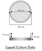
| Figure 1: Schematic representation of disks designed for these studies. They are 32mm in diameter with a 7. 5mm rim around the edge, and fit in 6-well culture plates used in the experiment. |
Since the predominant wear debris identified in periimplant tissues is ultrahigh molecular weight polyethylene (UHMWPE) from the acetabular liner, significant efforts have been directed towards improving the quality of the UHMWPE. Modifying the sterilization protocols to prevent oxidationinduced damage has enhanced the wear resistance of UHMWPE. Recent endeavors in cross-linking of UHMWPE have markedly improved the wear resistance of UHMWPE liners. 3, 5, 8 Laboratory hip simulators using cross-linked UHMWPE have demonstrated decreases in wear rates greater than 70% in comparison to those of conventional UHMWPE. Several variations of crosslinked liners have been approved by the FDA and are currently in use in the clinical setting.
Depending on the various processes used by the different companies, cross-linking of polyethylene alters the bonds between molecular chains, reduces crystallinity, alters the free radical content of the material and significantly influences the material and surface properties. 3 However, despite these changes in the physico-chemical characteristics of UHMWPE, we still do not know if the biological response to this material is altered. Thus the purpose of this study was to investigate the macrophage response to cross-linked UHMWPE and compare it to conventional UHMWPE surfaces. These investigations will provide essential insight into the inflammatory properties of these materials before the onset of wear and corrosion of the implant. This information can also be useful in directing future development of materials used in total joint replacements.
 |
|
|
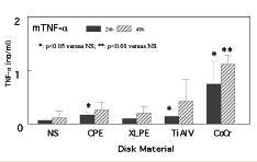
Figure 2: Murine TNF-a released by RAW macrophages exposed to lipped culture discs for 24 and 48 hrs. Experiments were conducted in triplicate and cytokine levels were determined using ELISA assays. Statistical comparisons were made between treatments and the control (NS) for that respective time. |
MATERIALS
AND METHODS
To carefully evaluate the response to the test materials, we designed
lipped culture disks that isolated the cells on the material and ensured
they did not contact other materials. The lipped culture disks were 32
mm in diameter with a 7. 5mm rim raised over the edge (See
Figure 1) . This design ensured that cells cultured on the disks
did not encounter other surfaces as well. Disks were fabricated from cross-linked
UHMWPE (XLPE) , conventional UHMWPE (CPE) , titanium-6aluminum 4 vanadium
alloy (TiAlV) , and cobalt-chrome alloy (CoCr) (Zimmer;Warsaw, IN) . The
raised lip provided containment of cell cultures with complete separation
from the standard tissue culture polystyrene wells in which they were
placed. Surface character and topographical profiles were determined for
each disk type. The UHMWPE disks were sterilized by plasma glow discharge
to preserve the cross-linking parameters, while the TiAlV and CoCr were
gamma-irradiated.
 |
|
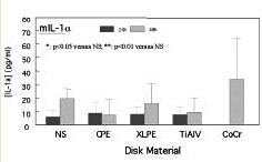
| Figure 3: Murine IL-1 a released by RAW macrophages exposed to lipped culture discs for 24 and 48 hrs. Experiments were conducted in triplicate and cytokine levels were determined using ELISA assays |
The murine macrophage cell line (RAW 264. 7, ATCC, TIB-71, Manassas, VA) was plated at 2. 0x10 6 cells per well in 2ml of media. The controls consisted of cells cultured on standard polystyrene tissue culture wells. Negative controls were non-stimulated (NS) cells, and cells stimulated by a lipo polysaccharide (LPS) were positive controls. Cells were plated in triplicate for each disk type and the experiment was repeated four times. Conditioned media was collected at 24h and 48h and analyzed for the presence of pro-inflammatory mediators IL-1 a and TNF-a . All collected media conditioned by the cell-disk interaction was analyzed using commercially available enzyme-linked immunosorbent assay kits (ELISA, R&D Systems, Minneapolis, MN).
The data was analyzed by one-way ANOVA and t -tests. An alpha level of 0. 05 was used as the criterion for statistical significance. Comparisons were made with respect to TNF-a and IL-1 a between stimulated cells and the control (NS) for that respective time period.
RESULTS
Non-stimulated murine macrophages released minimal levels of TNF-a at
24h (. 65 ng) , which increased at 48h (1. 13 ng) after culture on standard
6-well polystyrene tissue culture plates (See
Figure 2) . Samples stimulated with LPS demonstrated a 46-fold
increase in TNF-a secretion (29. 83 ng) at 24h and a 36-fold increase
(41. 79 ng) at 48h. Cells cultured on the polyethylene species were only
marginally more stimulated than NS cells. XLPE disks stimulated cells
to secrete 1 and 2ng of TNF-a at 24h and 48h, respectively, and CPE disks
stimulated cells to secrete 1.7 and 2.6 ng at 24h and 48h, respectively.
TiAlV samples were similarly nonstimulatory at 24h (1. 36 ng) but tripled
secretion to 4. 24 ng at 48h culture. In sharp contrast, macrophages cultured
on CoCr disks produced greater amounts of TNF-a for both the 24h samples
(7. 47 ng) and 48h samples (11. 14 ng).
Non-stimulated murine macrophages released 5. 95 pg of IL-1 a at 24h and 19. 41 pg at 48h (See Figure 3). Stimulation with LPS elicited a 7-fold increase at 24h (37. 41 pg) and a 100-fold increase at 48h (1908 pg) . In parallel with the TNF-a profile, both UHMWPE types did not significantly increase the release of IL-1 a from the non-stimulated level (the profile illustrates a range from 7 to 16pg of IL-1 a secretion for both polyethylene species-similar to that of the non-stimulated cells) . Similarly, TiAlV demonstrated very little stimulatory effect on the cells, which released 7. 6 and 9. 3pg at 24 and 48h, respectively. There was no IL-1 a detected in CoCr samples at 24h, but there was a large amount seen at 48h (33. 83pg) .
DISCUSSION
These studies suggest that there is no significant biological difference
between cross-linked UHMWPE and conventional UHMWPE. Since cross-linking
of UHMWPE acetabular liners is a recent development in biomaterials and
makes up a substantial portion of hip replacements on the market today,
it is important to document the immunological response of macrophages
to cross-linked UHMWPE surfaces. This will predict the initial response
of these cells to the introduction of a newly implanted prosthesis when
wear debris has not yet become a detrimental factor leading to osteolysis
and aseptic loosening.
The transformed murine macrophage cell line was used for its reproducibility and consistency in response. Although there are variations in the amount and type of cytokine released between human and mouse cells (for example, IL-1 a release is more prevalent in human macrophages rather than IL-1 ß) , there are definite correlations which can be taken from the overall response of these cells. Likewise, because the RAW macrophages are one of the established models used to study immunological response and are available to other researchers around the world, direct comparisons can be made with other materials and also with human monocytes.
Our studies have demonstrated that there is minimal murine macrophage response to XLPE and CPE disks. The cytokine profiles indicate that the cells maintained basal levels of TNF-a and IL-1 a . Using the positive control (LPS) demonstrates the broad stimulatory capacity of the cells. Using titanium-alloy and cobalt-chrome alloy disks permitted us to understand the general macrophage response to these two commonly used implant materials in comparison to UHMWPE. These studies illustrate that despite changes in molecular bonds, crystallinity and free radical content of polyethylene due to cross-linking, murine macrophages could not differentiate between XLPE and CPE surfaces. It is still not clear if murine macrophages would be able to recognize and respond differently to particles from the two PE species, where the cells would be in more intimate contact with the materials. These investigations are currently ongoing in our laboratory.
ACKNOWLEDGMENTS
We would like to acknowledge Dale Swarts (Zimmer, Inc. ) and William Clarke
(Zimmer, Inc. ) . This study was supported by a grant from Zimmer, Inc.
Mark J. Neavyn is a Research Assistant, Orthopaedics Research Laboratories, Massachusetts General Hospital, Boston, MA
Rajiv K. Sethi, BS is a Medical Student, Harvard Medical School. Harry E. Rubash, MD is a Professor in Orthopaedic Surgery, Harvard Medical School.
Arun S. Shanbhag, PhD is an Assistant Professor in Orthopaedic Surgery, Harvard Medical School.
Address correspondence to:
Arun S. Shanbhag, PhD
Department of Orthopaedic Surgery
Massachusetts General Hospital
54 Fruit Street
Boston, MA 02114