|
|
|
| Click here to visit our web site |
|
INTRODUCTION The physeal bar is a corollary of the focal articular cartilage lesion in proliferating cartilage. If small, the physeal bar is a tether that interferes with the kinetic program of the surrounding intact growth plate. Resection and replacement with inert fillers are the present solution, limited by the requirement that at least fifty percent of the growth plate must be intact to allow acceptable growth. Focal replacement of the resected physeal region with cartilage has been attempted experimentally, but it is unclear whether such transplanted cartilage provides cells that participate in growth. Furthermore, even if the transplanted cartilage had kinetic activity, it may not match that of the remaining physeal cartilage following untethering. An even larger challenge would be the engineering of an entire joint that was congenitally absent or severely damaged. Repair of congenital or acquired joint deformities requires the creation of compound structures with complex anatomical and functional features. Progress in musculoskeletal cell, developmental, and molecular biology, advances in in vitro histogenesis, and innovations in materials and manufacturing processes have not yet resulted in widespread clinical solutions to such devastating problems. RESEARCH PROGRAM Several means for delivering the viable chondrocytes were evaluated. Aliquots of a suspension of bovine articular chondrocytes (bACs) were inoculated into the distal femoral and proximal tibial devitalized chick chondroepiphyses with a dissecting microscope and a micromanipulator. Small foci of viable chondrocytes were identifiable with this method but not with the desired widespread penetration. Bovine chondrocytes were precultured with braided polyglactic acid suture for 5-28 days and were found to adhere to and permeate the interstices of that braided material. The suture was then passed into devitalized chick knees. Histologic analysis showed that viable bovine chondrocytes were present, predominantly at the outer surface of the suture where it interfaced with the devitalized chick matrix. In the next test, canals were created in devitalized chick chondroepiphyses either with a 2812-gauge needle or with an Er:YAG laser with an average of 15 canals per joint surface. A suspension of cells was placed onto the prepared surface for culture. Viable chondrocytes were found in the canals made either way. There was filling of the canal with some invasion of the devitalized matrix. This became more extensive with longer time in culture. In another series, canals were created by the physical means described. The chondroepiphyses were further treated with lytic enzymes (2. 5 mg/ml trypsin, 1 mg/ml hyaluronidase and 1 mg/ml collagenase) for only 10 minutes and re-lyophilized. The treated joint surfaces were rehydrated with a suspension of cells and were cultured for 7 and 21 days. There was enhanced infiltration of viable chondrocytes with the addition of enzyme pre-treatment. This was interpreted as a consequence of the enzymes’ rendering the matrix more penetrable. IN VITRO AND IN VIVO CHARACTERISTICS OF KNEE
CONSTRUCTS
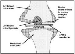
The most successful model we have developed to date uses porous collagen sponges to deliver the viable chondrocytes to the anlage (Figure 1). This phase of the program uses an experimental construct composed of an embryonic chick devitalized by lyophilization and viable chondrocytes isolated from neonatal bovine articular cartilage and delivered to the joint in porous collagen sponges (Figure 1). The sponges are meant to contain the cells in the desired location. These collagen sponges have been shown to support chondrogenesis (1), chondroinduction (2,3), osteogenesis (4), and in vivo compatibility (1,5). Constructs were engineered in vitro and their ectopic in vivo fate was examined. (6) Chimeric joints were made by affixing porous collagen sponges that contained bACs to opposing shaved articular femoral and tibial surfaces of devitalized embryonic (19 d) chick knees. Membranes of expanded polytetrafluoroethylene (ePTFE) were positioned between the femoral and tibial sponges. The constructs were cultured and subsequently transplanted into heterotopic sites in severecombined- immunodeficiency-disease (SCID) mice, chosen to avoid immune rejection of the bovine cells. The constructs were analyzed by histology at intervals in vitro and in vivo and by gene expression analyses for bovine markers. After 1 week in vitro, collagen sponges with bACs were adherent to the shaved articular surfaces of the devitalized chick joints. Subsequently, metachromatic neocartilage accumulated in the sponges. The bovine chondrocytes were the source of the neocartilage, as demonstrated by RT-PCR analysis of extracted RNA. Molecular analysis showed expression of bovine cartilage genes (collagen type II, aggrecan core protein, and aggrecan link protein) for the 3-week observation period. The ePTFE membranes were successful in maintaining a joint space between opposing sponges. Some constructs were cultured for 1 week before transplantation into dorsal subcutaneous pouches of 5-week-old immunodeficient mice. Chimeric joints exhibited dramatic changes in vivo. The bACs invaded the articular matrix of the devitalized knees. At 6 weeks, bovine neocartilage replaced much of the chick cartilage. At 8 weeks, bAC proliferation and neocartilage formation transgressed the scaffolds and created a synchondrosis around the joint. This duration is considered to be excessive in this unloaded site. Seeded bACs repopulated the devitalized chick scaffold and ePTFE membranes maintained knee joint spaces. ENABLING TECHNOLOGIES Proliferation and matrix production were shown be exuberant and, in this immobilized, unloaded system, often resulted in synchondrosis across the two sponges. This was effectively prevented by insertion of ePTFE membranes between the sponges, but may not be needed in mobile and loaded sites. Bovine neocartilage penetrated into the devitalized chick articular cartilage in vitro and in vivo. In SCID mice, the bovine neocartilage replaced the chick scaffold at 6 weeks, but transgressed the construct at 8 weeks; this duration is considered to be excessive in this unloaded site. The in vivo subcutaneous environment in SCID mice was conducive to cartilage formation in joint constructs. Because of the immunodeficiency of these test animals, it is possible, albeit unlikely, that host responses to bioengineered joints may involve an inflammatory element that would require modulation. Recently, chondrogenesis has been enhanced in collagen sponges in vitro by several means. A small amount of hyaluronan (2% of dry weight) that had been incorporated into threedimensional collagen scaffolds enhanced chondrogenesis, but a greater amount was inhibitory, apparently due to excessive filling of the porous nework. (7) The results with the composite hyaluronan/collagen sponge show the potential for modifying scaffolds to improve production of engineered cartilage for in vivo applications. Mechanical forces have also been shown to modify chondrogenesis in vitro. With a novel fluid pressure culture system, 2.8 MPa fluid pressure applied in either a constant or cyclic manner stimulated synthesis of cartilage specific proteoglycans by chondrocytes cultured in 3-dimensional collagen sponges. (8) Optimal matrix production may be achieved in bioengineered joints using hyaluronan/collagen sponges, preculture with fluid pressure, as well as with supplemental growth factors. CLINICAL RELEVANCE SUMMARY ACKNOWLEDGEMENTS Julie Glowacki, PhD is Director, Skeletal Biology Laboratory, Brigham and Women's Hospital and an Associate Professor of Orthopaedic Surgery, Harvard Medical School. David J. Zaleske, MD is Professor of Orthopaedic Surgery, Children's National Medical Center, Washington, DC. Address correspondence to: |
|
Print Manuscript • View References • Download PDF version • Close window |