|
|
|
| Click here to visit our web site |
|
INTRODUCTION Based on mechanobiological means and information, we intend to improve upon the repair or replacement of damaged tissue. The biology of chondrocytes in response to physical, physicochemical, and mechanical stimuli are still unclear. This article focuses on my current work: 1) mechanosignal transduction and mass transfer stimulated with hydrostatic pressure and/or strain to elucidate the signal cascade at the cellular and tissue level, and 2) the development of an in vitro tissue processor for cell-based therapy, applied techniques and instruments used in mechanobiological investigation. MECHANOSIGNAL TRANSDUCTION Cellular activities – such as proliferation, phenotypic expression, and metabolic activities – are altered due to mechanical changes. Mechanical signals are transduced through intracellular second messengers, such as cAMP, inositol phosphate, and calcium ions. Intracellular calcium is a widely used marker which can be measured using a fluorescent indicator. Technical difficulties in measuring intracellular calcium concentration ([Ca]i) have included the transient nature of changing calcium concentrations and the application of hydrostatic pressure to live cells using an optical instrument. The author developed a culture chamber in which a sapphire glass windows allows for an inverted microscope to non-invasively measure [Ca]i using a laser confocal microscope. The dynamic [Ca]i in chondrocytes may be measured using a fluorescent calcium indicator (X-rhod-1) and a laser confocal microscope system after applying pressure. This system allows hydrostatic fluid pressure application while minimizing fluid shear stress due to fluid flow and octahedral shear stress due to cell deformation. Articular cartilage has longitudinally heterogenous histology, which can be divided into three zones: surface, middle, and deep. Traditionally, a homogenous pool of chondrocytes has been used in most experimental settings. Differences in cell and matrix components due to age, loading history, thickness, and varying pathophysiological conditions were ignored. In later experiments, the focus was placed on the loading history of each cell as cellular behavior and calcium concentration were measured.
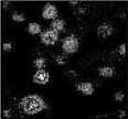
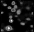
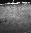
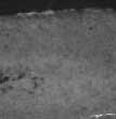
In our experimental system, cartilage from each zone was harvested from the weight-bearing site of a bovine forelimb condyle. Cells were isolated enzymatically from the cartilage pieces and used for signal transduction experiments. The calcium indicator was loaded prior to pressure application. Hydrostatic fluid pressure was tested varying from 0 – 3 MPa and 0 - 0.025 Hz. Pressure was applied for 5 min with varying conditions. After releasing the pressure, fluid flow was stopped and images were acquired at atmospheric pressure. Imaging was scanned for 800 nsec at 10 sec intervals for 5 min. Relative fluorescent intensity was measured as the summation of the cells in a region of interest, 8 cells to one frame. [Ca]i increased in chondrocytes harvested from all three zones. (3) [Ca]i peak values of middle zone cells was two times higher than surface and deep zones (Figure 1). In order to avoid involvement of cytoskeletal motion, cytochalasin B, which depolymerizes actin filaments of cytoskeleton, was added prior to pressure application. (4) Even without cytoskeletal effects, [Ca]i increased after releasing pressure, but the peak value was less than with cytoskeleton. This indicates that the cytoskeleton contributes to calcium signaling to some degree. This project has been expanded to determine the location of the source of calcium and further characterize the signal cascade at the subcellular level. Most likely, pericellular matrix components should be included in a capacity of boundary water under pressure. MASS TRANSFER IN CARTILAGE Our hypothesis was that fluid exudation from the articular surface due to joint loading is minimized. We harvested cartilage discs from weight bearing sites of the forelimbs of 2 – 3 week old calves. These discs were incubated with fluorescent tracers, rhodamine-dextran or fluoresceinisothiocyanate (FITC)-dextran, each with a defined molecular weight. The discs were incubated for 6 and 12 hours. The cartilage was frozen immediately after samples were harvested. Sections were cut 20 µm thick. Fluorescent intensity was acquired using a laser confocal microscope. Highest fluorescent intensity was detected in the middle zone. However, less intensity was detected from the longitudinal side of the disc (Figure 2).
From an engineering perspective, it is important to consider that once chondrocytes accumulate their own extracellular matrix, the permeability of the cell/construct decreases. We also have to consider molecular sheaving by the tissue. We found that FITC-dextran diffused into cartilage discs primarily from the articular surface and from the subchondral side, but far less from the site of the longitudinal cut (unpublished data). We suspect that the physicochemical environment of this tissue may influence cells to restrict molecular sheaving. This is a very important concept to consider when designing three-dimensionally organized articular cartilage. CELLULAR AND TISSUE ENGINEERING FOR CELLBASED
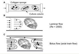
THERAPY MEDIUM PERFUSION We tested a number of different cell types using a medium perfusion system designed by the author and his colleagues. We found that medium perfusion enhances viability and hematopoiesis in murine marrow cells (5), osteogenesis in murine osteosarcoma (6) and oral mucosa (7). However, chondrocytes did not benefit in the same manner. (8) Each perfusion culture system has different features. Thus, we must carefully choose a system and optimize conditions for the target cell type. Fluid shear stress generated by fluid flow is a critical factor in cardiovascular cells such as endothelial cells. In cartilage, on the other hand, the interstitial fluid flow rate is extremely small. When we designed our perfusion culture system, we calculated the Reynold’s number and defined laminar flow (Re << 2000). We did not intend to mimic interstitial fluid flow. With our perfusion system, cell constructs are freely suspended in the column, thereby creating obstacles to the fluid flow. Theoretically, this flow, called Bolus flow (axial-train model, Figure 3), accelerates diffusion and convection of molecules, subsequently increasing nutrient supply as well as releasing products such as unbound, free extracellular matrix. Construct materials must be chosen depending on their permeability, because we must take into account not only the construct material’s properties, but also the effect of matrix accumulation on the cells themselves. By understanding these factors, we can control the fluid flow rate for the target cells and allow them to accumulate their own matrix.
PRESSURE/PERFUSION CULTURE SYSTEM TISSUE ENGINEERING PROCESSOR In addition, this tissue processor allows us to attach a “compression and stretch” apparatus to further investigate the mechanobiology of skeletal tissue. We plan to apply other physical forces and alter physicochemical conditions for these and other target tissues.
ACKNOWLEDGEMENT Suichi Mizuno, PhD is an Instructor in Orthopaedic Surgery, Harvard Medical School, Brigham and Women’s Hospital. Address Correspondence to:
|
|||||||||||||||||||||||||||||||
|
Print Manuscript • View References • Download PDF version • Close window |