|
|
|
| Click here to visit our web site |
|
INTRODUCTION We have used an aspect of nanotechnology to improve the performance of acrylic bone cement. We have developed a new nanocomposite polymethyl methacrylate (PMMA) bone cement with improved fatigue properties for application in the fixation of total joint replacement prostheses. All commercial cements contain approximately 10% weight of 0.5-3 µm size barium sulfate or zirconium oxide particles. These particles are radiopacifiers, which enable orthopedic surgeons to monitor the implanted cement using x-ray radiographs. This is necessary since cements are known to fail due to fracture, resulting in implant loosening and requiring revision surgery. Early fracture of cement has led to several studies (1-4) that focus on the possible reasons for cement fracture. Topoleski et al1 and Demian et al (2) have shown that agglomeration of radiopacifier particles is associated with cement fracture. Improvement in their dispersion can therefore lead to increase in fracture toughness of bone cement. In these studies, the 1-3 µm size barium sulfate particles were substituted with 100 nm size barium sulfate particles in a commercial bone cement. It was hypothesized that smaller radiopacifier “nanoparticles” could be more uniformly dispersed in the cement and would thus have the potential to improve its fracture toughness. Recent advances in nanotechnology have led to the commercial availability of “nano-sized” barium sulfate powder particles, making it possible to explore the effect of particle size on dispersion and fracture toughness of cements. Our preliminary fatigue tests showed that the “nanocomposite” cement had a substantially longer fatigue life than the currently used “microcomposite” cement. MATERIALS AND METHODS Fatigue tests were performed on an Instron 8511 servohydraulic testing machine in the laboratory of Professor Lisa Pruitt, Department of Mechanical Engineering, University of California at Berkeley. Prior to testing, the specimens were soaked in room temperature water for 2 days. During testing, the specimens were maintained at 37oC using a recirculating water bath. The pre-notched specimens were fatigued in load control with an R-ratio (minimum load to maximum load) of 0. 03 and stress amplitude of 15 MPa at a frequency of 2 Hz. Specimens that failed at the grips were discarded. Sample size ranged from 6-10. A JEOL 6320FV field emission low voltage scanning electron microscope (LVSEM) at the Center for Materials Science and Engineering at the Massachusetts Institute of Technology, Cambridge, MA was used to examine all fracture surfaces. In addition, ultra-small angle x-ray scattering (USAXS) at the UNICAT synchrotron x-ray beamline(5) of the Advanced Photon Source, Argonne National Laboratory, Argonne, IL was used to quantify the dispersion of radiopacifier particles in the microcomposite and nanocomposite PMMA cements. Sheets of 0.5mm thickness of each type of cement were subjected to USAXS using 10 kEV x-rays and a beam cross-sectional area of 2mm x 0.6mm. The x-ray scattering curves were analyzed using Porod’s law(6) to characterize the surface area to volume ratio (specific surface area) of dispersed radiopacifier particles as well as voids within the PMMA matrix. A reduction in the specific surface area of radiopacifiers detected by USAXS would imply particle agglomeration. Thus USAXS can be used for quantitative comparison of welldispersed and poorly dispersed radiopacifier particles within the cement. USAXS is advantageous in comparison to imaging methods in that the experiment estimates the average specific surface area over a relatively large sampling volume of approximately 1 mm3. RESULTS
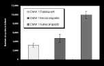
Fatigue tests showed that the nanocomposite CMW1 cement had a fatigue life of over a 100% longer than that of the microcomposite CMW1 cement containing regular barium sulfate (ANOVA-Bonferroni method, p<0.05), as shown in Figure 1. There was no statistically significant difference in the fatigue life of the radiolucent and microcomposite CMW1 cements for the number of samples tested.
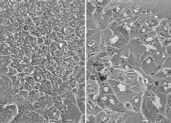
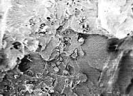
LVSEM observation of fracture surfaces revealed the presence of 0.5-3.0 µm size barium sulfate particles in the microcomposite cement (Figure 2). In addition, LVSEM revealed uniform dispersion of the nanometer-sized barium sulfate, a result that is likely due to the addition of sodium citrate as an anticoagulant. There was a higher concentration of features associated with fracture in the nanocomposite cement (Figure 3). These were confined to the regions containing the barium sulfate nanoparticles, and were not observed in the regions of the pre-polymerized powder. X-ray radiographs of tensile specimens confirmed that both composite cements were radiopaque.
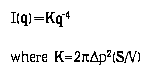
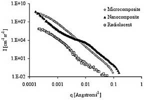
USAXS scattering curves were obtained by plotting the scattered intensity (I) vs (q) as shown in Figure 4, where: such that ¥è equals one half of the scattering angle and ¥ë is the wavelength of x-rays (=2. 38 Angstrom). USAXS revealed a substantial scattering intensity due to the presence of both voids (radiolucent cement) as well as due to barium sulfate particles (Figure 4). The region of the scattering curve at q > 0. 008 [Angstrom-1] was curve fitted to a Power law function which is:
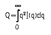
such that ¥ñ is the electron density difference between barium sulfate (or voids, in the case of radiolucent cement) and PMMA and (S/V) is the specific surface area of the scattering entity (radiopacifier or voids). The scattering invariant, Q, defined by the following equation was calculated for all cement samples using the area under the scattering curve, q2I(q) versus q:
The invariant for an angular range of 0-qmin was calculated by fitting the low q region of the scattering curve using Guinier¡¯s Law (6) and for qmax ¨C ¡Þ using Porod¡¯s Law. The specific surface area can then be defined as: S/V=¦Ð (K/Q). Porod analysis showed that the ratio of the specific surface area for the nanocomposite and microcomposite cements respectively was 9.16 (Table 1).
DISCUSSION This study also showed that USAXS is effective in quantitatively measuring the specific surface area of barium sulfate radiopacifiers dispersed within PMMA bone cement. It is expected that for the same volume (or weight) fraction, the nanometer size particles must have a total specific surface area that is 10 times larger than the micrometer size particles in bone cement since there would be a 1000 times more particles of 1/10th of the diameter. The value of 9.16 is in excellent agreement with this calculation, considering that the inherent particle size distributions can alter the ratio of specific surface area. Agglomeration of particles would result in a reduction in specific surface area. USAXS therefore showed that both microcomposite and nanocomposite cement had relatively well-dispersed radiopacifier particles. In addition, USAXS was able to provide the specific surface area of voids. This technique could be used to guide in the development of new mixing protocols with the objective of reducing voids since these flaws can also reduce the fracture toughness of cements. LVSEM confirmed the quantitative analysis provided by USAXS measurements by revealing fracture surfaces where particles were uniformly dispersed throughout the PMMA matrix. The large concentration of features associated with fracture of the nanocomposite suggests that there was extensive crack tip blunting, probably due to the large number of barium sulfate nanoparticles encountered by the propagating crack. In conclusion, the fatigue performance of acrylic cements can be substantially improved by a uniform dispersion of barium sulfate nanoparticles. However, there must be more rigorous testing under conditions that simulate in vivo loading and biological environments before these nanocomposite cements may be implemented in clinical practice. ACKNOWLEDGMENTS
Dr. Wolfgang Fitz is an Attending Surgeon at Brigham and Women’s Hospital and Instructor of Orthopaedic Surgery at Harvard Medical School. Dr. Andreas Gomoll is a Resident in the Harvard Combined Orthopaedic Surgery Program. Mary Beth Turell is a Research Assistant in the Orthopaedic Research Laboratory, Brigham and Women’s Hospital. Dr. Richard D. Scott is an Attending Surgeon at Brigham & Women’s Hospital and Professor of Orthopaedic Surgery at Harvard Medical School. Dr. Thomas S. Thornhill is Orthopaedist-in-Chief at Brigham & Women’s Hospital and John B. and Buckminster Brown Professor of Orthopaedic Surgery at Harvard Medical School. Address correspondence to: |
|||||||||||||||||||||||||||
|
Print Manuscript • View References • Download PDF version • Close window |