|
|
|
| Click here to visit our web site |
|
INTRODUCTION BACKGROUND The idea that even invisible scaphoid fractures can fail to heal has created a great deal of concern among physicians treating wrist injuries. The standard treatment of patients with “snuffbox tenderness” after a fall is substantial, including a minimum of two weeks of immobilization, repeat examination and radiographs and even a bone scan. (2) Attempts to explain the substantial nonunion rate of immobilized fractures of the scaphoid have focused on its meager blood supply and the mechanical stresses to which it is subjected. (2,3) Known fractures are immobilized for a minimum of 10 weeks, often much longer. This cast usually includes the thumb and often extends above the elbow. (4) NEWER CONCEPTS
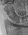
An alternative interpretation of these observations may also have merit. Few studies of scaphoid fractures have accounted for displacement,(1) despite the fact that displacement may be the single most important factor associated with healing problems. (5) The five to ten percent nonunion rate in most series could simply be a reflection of the incidence of displaced fractures. Non-displaced fractures of the scaphoid probably heal if adequately protected. As evidence in favor of this, a protocol underway in London, Ontario has treated over 50 patients with CT-confirmed non-displaced fractures of the scaphoid in a below elbow thumb spica cast for 10 weeks with no healing problems (personal communication, Graham King, MD). This has been our experience as well. Because scaphoid fractures occur in young, active people, nonunion after a radiographically invisible fracture may also reflect reinjury of an incompletely healed fracture in a person who returns to unprotected activity too soon. The initially undisplaced fracture may displace in a subsequent injury and ultimately fail to heal (Figure 1). Although some advocates of routine operative treatment of scaphoid fractures cite the five to ten percent nonunion rate of cast-treated fractures as unacceptable, operative treatment of non-displaced fractures should be considered primarily for its short-term benefits including avoidance of cast wear and earlier return to athletics or occupation. In a recent prospective, randomized comparison of operative versus nonoperative treatment of non-displaced fractures of the scaphoid, cast-treated patients lost very little motion or strength when compared to operatively treated patients and there were no healing problems in either group. (6) The advent of small cannulated screws has facilitated percutaneous fixation of the scaphoid. However, the screws and the tools used to insert them are delicate and can be technically difficult to use; caution is warranted. (7) There is general agreement that displaced fractures require operative treatment for realignment and stable fixation. (1,2,5) This can sometimes be accomplished percutaneously with wrist arthroscopy and image intensification used to monitor the reduction. Some nonunions are stable and well aligned. Many of these are fibrous unions with an intact cartilage or fibrocartilage shell when exposed operatively. It has recently been suggested that these stable, fibrous nonunions can be treated with percutaneous screw fixation and that neither open debridement nor bone grafting are necessary. (8) Computed tomography or arthroscopy may be necessary to distinguish stable fibrous nonunions from atrophic or synovial nonunions. The concept merits further study. CURRENT PROJECTS MRI has been suggested as a diagnostic modality for rapid and accurate triage of suspected fractures of the scaphoid(9); however, MRI has several important drawbacks. MRI is expensive and not readily available in most hospitals. In addition, interpretation of the scans is not straightforward. Due to the small size of the scaphoid and absence of thick, distinct cortices, fractures and other changes such as “bone bruises” can be difficult to distinguish.
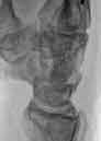
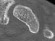
Computed tomography is more readily available and less expensive in most hospitals. In fact, it is already commonly used in Europe for the triage of suspected fractures of the scaphoid. We are currently using a protocol approved by the Human Research Committee to evaluate the use of computed tomography for the triage of suspected scaphoid fractures. Images obtained in planes defined by the long axis of the scaphoid are much easier to interpret than transverse scans of the wrist10 (Figure 2). To obtain sagittal plane images along the axis of the scaphoid, the patient lies prone with the injured arm overhead and the hand flat on the gantry. The forearm crosses the gantry at a 45-degree angle and the axis of the scaphoid corresponds roughly with the radially abducted thumb. To obtain coronal plane images the forearm is supinated 90 degrees. Another useful aspect of CT scanning is the ability to identify other radiographically occult injuries that can explain the patient’s pain, such as a non-displaced fracture of the distal radius or trapezium. (11) In our experience, surface rendered three-dimensional computed tomography reconstructions have not been helpful.
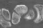
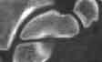
Unfortunately, computed tomography cannot reliably distinguish a non-displaced fracture from a vascular channel or other line seen on computed tomography (Figure 3). In a comparative study of non-fractured scaphoids from our suspected scaphoid protocol and fractured scaphoids from another protocol (described below), we found that nondisplaced fractures are often unicortical and may only be seen on one or two slices in each plane. The normal scaphoid has linear opacities on computed tomography, probably vascular channels, that are difficult for both radiologists and orthopaedic surgeons to distinguish reliably (sensitivity 94%, specificity 90%). (11) We have concluded from our preliminary studies that there is, as of yet, no ideal diagnostic test for the triage of suspected scaphoid fractures. NON-DISPLACED FRACTURES OF THE SCAPHOID: OPERATIVE
VS. NON-OPERATIVE TREATMENT We have a prospective protocol comparing operative and non-operative treatment and have learned several things simply by attempting this study. First of all, it has been our experience that patients are extremely reluctant to be randomized between operative and non-operative treatment. Some authors have even argued that prospective randomized trials are unethical because they interfere with the doctor-patient relationship. (12) Bond and co-authors had difficulty enrolling patients, ultimately enrolling only one-third of eligible patients in what would otherwise be considered favorable circumstances for performing such a trial. (6) Our Human Research Committee would not allow a surgeon-randomized trial, instead insisting that the study be a non-randomized trial, or “open label,” meaning that patients are fully informed of all options and able to choose their treatment. When we first initiated the trial, the non-operative treatment protocol began with six weeks of above elbow thumb spica cast wear. As patients with this injury tend to be young and active, most dread prolonged cast wear. Among the first eight patients enrolled in the trial, only one elected cast treatment. Since changing the non-operative protocol to below elbow casting from the outset, we have had better luck enrolling patients in the non-operative arm of the study; however, operative treatment is still favored by the majority of patients. Our results to date confirm the findings of Bond and colleagues, with no significant differences in outcomes between treatment groups. COMPUTED TOMOGRAPHY ANALYSIS OF THE SCAPHOID
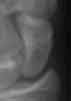
In one study, we looked at the placement of a partially threaded cannulated screw into the scaphoid using a volar insertion point. Successful volar insertion includes placement of the tip of the screw in the center of the proximal pole with all of the threads of the screw in the distal fracture fragment. The challenge is to work around the trapezium, avoid volar cutout of the screw, and avoid perforating a joint surface. Our analysis suggested that these challenges are best met by using a relatively radial starting point (Figure 4). In addition, the most likely area for perforation of a joint surface is the dorsal radial surface of the scaphoid. (13) This area is best evaluated on a partially pronated anteroposterior view of the wrist. In another study we investigated the effectiveness of computed tomography in evaluating scaphoid deformity by measurement of intra-scaphoid angles, dorsal cortical angles, and height to length ratios. Our studies have confirmed the findings of other investigators that these measurements vary substantially between different observers and even with the same observer over time. In addition, the obliquity of the scanning angle (which is set in a relatively arbitrary manner), can have a substantial affect on these measurements. Our preliminary conclusions are that, while computed tomography is the best method for evaluating and visualizing deformity, the techniques used to quantify the deformity are imprecise, unreliable, and vary with technique. (13)
CONCLUSIONS David Ring, MD is Instructor, Orthopaedic Surgery, Harvard Medical School, Hand and Upper Extremity Service, Department of Orthopaedic Surgery, Massachusetts General Hospital Jesse B. Jupiter, MD is Professor, Orthopaedic Surgery, Harvard Medical School, Chief, Hand and Upper Extremity Service, Department of Orthopaedic Surgery, Massachusetts General Hospital Address correspondence to: |
||||||||||||||||||||||||||||||||||||
|
Print Manuscript • View References • Download PDF version • Close window |