HOJ HOME | Chiefs Reports | Osgood Day | Cartilage Regeneration and Repair, Where Are We?
A Harvard Orthopaedic Presence in China
|
Scientific Articles | Alumni

visit our website at: www.gensci.bc.ca
| click here to view full page ad
Hip Dysplasia in the Adult:
Joint Preserving Methods to Prevent or Treat Osteoarthritis

Michael B. Millis, MD • Stephen B. Murphy, MD

The Children's Hospital and Beth Israel Deaconess Medical Center

          Dysplasia is the single most common etiology of osteoarthritis of the hip.1-4 Recent work has clarified factors prognostic for osteoarthritis in the mature dysplastic hip allowing identification of those patients at risk for osteoarthritis before radiographic arthrosis is present.5 Since 1957, when Salter carried out the first innominate osteotomy and introduced the era of acetabular redirection, increasingly powerful techniques of acetabular realignment have evolved to allow correction of the acetabular obliquity and instability that have been closely associated with the development of arthrosis. Over more than 25 years, a major clinical experience has been gained within the Harvard Orthopaedic Program with acetabular redirection osteotomies to treat the mature dysplastic hip. Fifty spherical Wagner-type acetabular osteotomies were performed between 1981 and 1992 and 275 Bernese (or Ganz-type) periacetabular osteotomies have been performed since 1991. Computed tomography (CT)-based analyses of the acetabular deformity in developmental dysplasia have allowed quantification of pathological mean joint contact pressures; simulation of the reduction in pathologic joint contact pressures expected from appropriate acetabular realignments; and confirmation of these contact pressure reductions through analysis of postoperative CT scans. With pain and congruous acetabular dysplasia representing the indications for surgery, relief of pain, improvement of limp, normalization of acetabular alignment, and improvement in bone structure has been nearly universal, particularly with the contemporary abductor-sparing direct anterior surgical approach.

Background

The treatment of osteoarthritis of the hip has been an interest of Harvard orthopaedists since Smith-Petersen first developed the anterior ilio-femoral approach and subsequently employed it to establish and refine the important technique of mold arthroplasty during the first third of the 20th century. Harris and Stulberg, in the early 1970's first recognized the strong association of developmental deformities of the hip with the occurrence of osteoarthritis.2 John Hall, also in the early 1970's, advanced the practice of joint preserving hip surgery when he brought to Boston the techniques of innominate osteotomy and Chiari osteotomy to treat acetabular dysplasia in the adolescent and young adult.

          Increasing recognition of hip dysplasia as the single most common etiology of osteoarthrosis of the hip - certainly in women and perhaps in men as well - has led to the foundation of strong clinical and research interests in hip dysplasia at The Children's Hospital, Beth Israel Deaconess Medical Center, and Brigham and Women's Hospital. In 1978-1979, Michael Millis spent an AO Fellowship with Heinz Wagner in Germany, importing the techniques of complex inter-trochanteric osteotomies and spherical osteotomies to treat the deformities of dysplasia in the young mature hip. Robert Poss spent a sabbatical in Europe in the early 1980's, also learning osteotomy techniques from Wagner, as well as from Maurice MŸller and Renato Bombelli. Stephen Murphy spent a fellowship with Maurice MŸller and Reinhold Ganz.

           Following these European experiences, Millis, Murphy, and Poss each made major commitments to joint preserving surgery. To complement and improve the clinical practice - which now comprises more than 300 complex acetabular osteotomies carried out over a period of more than 18 years - several parallel research efforts have contributed to the understanding of the pathomechanics present in hip dysplasia, as well as suggesting ways to improve the already impressive clinical results.


NEXT PAGE | TOP OF PAGE | HOJ HOME
Chiefs Reports | Osgood Day | Cartilage Regeneration and Repair, Where Are We?
A Harvard Orthopaedic Presence in China
|
Scientific Articles | Alumni


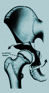
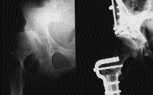
Operative Techniques for Joint-Preservation in the Mature Dysplastic Hip: Recent Refinements

          Hip arthroscopy has been employed extensively by Dr. Murphy for both surgical decision making in the hip with some arthrosis, in whom a decision must be made between joint preservation and joint replacement, as well as for removal of loose bodies and debridement of labral lesions.
The greatest recent advances in surgical joint preservation techniques for the dysplastic hip have involved the use of acetabular redirection.3, 4, 9-13 A satisfying clinical experience was gained using the spherical acetabular osteotomy (as learned by Drs. Millis and Hall from Professor Heinz Wagner) - an elegant procedure that achieved redirection using large spherical chisels. Although our clinical results were very good14, the procedure required an extensive anterior approach with reflection of the abductor muscles off of the iliac crest and there was some degree of permanent abductor dysfunction in a minority of hips. In addition, arthrotomy to deal with intra-articular pathology was believed to be unwise because of concern for the blood supply to the thin acetabular fragment, and was therefore not performed.

          With the recognition of the acetabular rim syndrome by Klaue and colleagues15, it has become desirable to perform anterior arthrotomy in association with redirection osteotomy in order to deal with the intra-articular pathology frequently present in adult dysplastic hips. In 1991, Millis and Murphy began employing the Bernese periacetabular osteotomy9 in selected patients in whom arthrotomy seemed indicated because of clinical signs of labral tears. (Figure 1) After a few initial cases done through a modified Smith-Petersen approach, the entire subsequent series of more than 250 hips treated by Bernese periacetabular osteotomy at Harvard has been performed through an exposure which spares the origin of the abductor musculature.16

Figure 1: The so-called Bernese, or Ganz periacetabular osteotomy is a powerful means of reorienting the dysplastic acetabulum.

The Direct Anterior Approach for Periacetabular Osteotomy

           Our modification of the operative exposure for Bernese periacetabular osteotomy, the so-called direct anterior approach16, combines the best features of both the Smith-Petersen and the ilio-inguinal exposures of Letournel. The development of this exposure was guided by the following principals: 1) the exposure must be sufficiently extensile to allow safe redirection of the acetabulum employing the osteotomy cuts described by Ganz and colleagues9; 2) it should allow anterior arthrotomy to deal with intra-articular pathology; and 3) it should avoid dissection of the abductors off the iliac crest with its related abductor morbidity. The direct anterior approach involves use of the medial half of the Smith-Petersen approach, with dissection between the sartorius and tensor fascia lata, as well as osteotomy of the anterior superior spine to relax the sartorius. The second window of the ilio-inguinal approach medial to the psoas and the femoral nerve, can be used to allow direct visualization of the deep medial cuts of the Bernese osteotomy along the medial wall of the pelvis. The entire operation can be performed lateral to the psoas, but medial to the ilium, in most patients if the surgeon is experienced; but the second window is used in most large muscular patients.

           A large clinical research project involving cybex testing of abductor function both before and after acetabular osteotomies carried out through various surgical approaches has confirmed the clear superiority of the abductor-sparing direct anterior approach in terms of the rapidity and completeness of recovery of abductor function following surgery.17 In fact, the group of patients operated on through the Smith-Petersen approach rarely achieved return to their levels of preoperative abductor strength over periods of observation as long as 24 months.


NEXT PAGE | TOP OF PAGE | HOJ HOME
Chiefs Reports | Osgood Day | Cartilage Regeneration and Repair, Where Are We?
A Harvard Orthopaedic Presence in China
|
Scientific Articles | Alumni


Clinical and Radiographic Results of Periacetabular Osteotomy in the Treatment of Acetabular Dysplasia in the Mature Hip
          Since 1991, more than 275 mature dysplastic hips have been treated with Bernese periacetabular osteotomy at Children's Hospital, the Beth Israel Hospital, and the Brigham and Women's Hospital. (Figure 2) To date, the average age of the patients is 27 years, the oldest being 55 years of age. Most of these hips had congruous dysplasia and little or no arthrosis, although approximately 25 hips had grade II or grade III arthrosis by the Tonnis scale. All of the hips treated with periacetabular osteotomy had pain preoperatively, and most patients limped. Most patients had an excellent range of motion preoperatively. Many patients older than 25 had symptoms suggestive of labral pathology in the form of intermittent giving way of the hip or episodes of painful locking. All patients had pathologically small lateral and anterior CE angles, with the mean in this group for both anterior and lateral CE angles being about +2¡. About 25% of patients older than 25 years of age have had labral pathology noted at arthrotomy - usually anterior labral tears which were debrided.

Figure 2: We have performed over 275 Bernese osteotomies in adolescents and adults with dysplastic hips.

A: This patient had bilateral hip dysplasia.

B: The right hip required varus intertrochanteric femoral osteotomy in addition to acetabular redirection.

C: This radiograph taken after healing and hardware remove demonstrates the improvement in joint orientation - with consequent decrease in contact pressures - that can be obtained.

          In a subset of 100 hips which have been followed for two years or more (maximum eight years), we have observed almost universal improvement in clinical function and excellent radiographic results. Mean post-operative anterior and lateral center-edge angles have been about 20 degrees. Subsequent total hip arthroplasty was required in six hips with preoperative grade III arthrosis when periacetabular osteotomy failed to relieve symptoms. Most of the remaining patients have experienced complete pain relief and are free from limp. Average time on two crutches was approximately two months. No external immobilization has been required. Approximately 15 percent of patients have had simultaneous proximal femoral osteotomy in the form of intertrochanteric or trochanteric osteotomy. (Figure 3)

           Subsequent surgeries performed, other than the six total hip replacements, have included two intertrochanteric osteotomies performed for residual subluxation or rotational malalignment in the proximal femur, as well as plating and grafting of one symptomatic ischial non-union and excision of anterior heterotopic bone from one muscular male athlete.

Figure 3: Bernese periacetabular osteotomy can be used to treat severe dysplasia provided that redirection results in congruent articulation of the femoral head with the acetabulum.

A: This patient had severe dysplasis of the right acetabulum and femur with subluxation of the hip.

B: Redirection of both the acetabulum and femur created more favorable mechanical conditions at the hip joint while maintaining congruency of the articular surfaces.


NEXT PAGE | TOP OF PAGE | HOJ HOME
Chiefs Reports | Osgood Day | Cartilage Regeneration and Repair, Where Are We?
A Harvard Orthopaedic Presence in China
|
Scientific Articles | Alumni


Conclusions

         The importance of acetabular dysplasia in the pathogenesis of many cases of osteoarthritis of the hip is clear, as are the radiographic criteria for identifying the patients at risk. Our clinical and research experience has led to continued improvement in our clinical results, in terms of both the completeness of the corrections achieved as well as the reduced perioperative morbidity for our patients. Current efforts focus on the identification of patients appropriate for treatment earlier in the disease process; determination of optimum intraoperative placement of the osteotomized acetabulum; and extended follow-up analyses to better characterize operative indications for joint preservation versus joint replacement and to determine optimal treatments of various pathologies of the acetabular rim.

Acknowlegements

         The authors express their deep gratitude to Dr. John Hall, Professor Heinz Wagner, Professor Maurice MŸller, and Professor Reinhold Ganz. We also recognize the important contributions of our colleagues, Dr. Robert Poss, Dr. John Hipp, Ph.D., and the support given our work by the AO Foundation and the Orthopaedic Research and Education Foundation. We also appreciate the contributions of the nurses, physical therapists and other health care colleagues at our institutions.

Michael B. Millis, MD is an Attending Surgeon at Boston Children's Hospital and Associate Professor of Orthopaedic Surgery at Harvard Medical School.

Stephen B. Murphy, MD is an Attending Surgeon at Beth Israel Deaconess Medical Center and Clinical Instructor in Orthopaedic Surgery at Harvard Medical School

Address for correspondence:
Michael B. Millis, MD; Department of Orthopaedic Surgery; The Children's Hospital; 300 Longwood Avenue; Boston, MA 02115
e-mail: millis@a1.tch.harvard.edu

References
1. Aronson J. Osteoarthritis of the young adult hip: Etiology and treatment. AAOS Instructional Course Lectures. St. Louis: Mosby, 1986.
2. Harris WH. Etiology of osteoarthritis of the hip. Clin Orthop 1986;213:20-33.
3. Millis MB, Poss R, Murphy SB. Osteotomies about the hip in the prevention and treatment of osteoarthritis. J Bone Joint Surg 1995;77A:426-437.
4. Poss R. Current concepts review: The role of osteotomy in the treatment of osteoarthritis of the hip. J Bone Joint Surg 1984;66A:144-151.
5. Murphy SB, Ganz R, Muller ME. The prognosis in untreated hip dysplasia: factors predicting outcome. J Bone Joint Surg 1995;77A:985-989.
6. Hipp JA, Sugano N, Millis MB, Murphy SB. Planning acetabular redirection osteotomies based on joint contact pressures. Clin Orthop In Press.
7. Millis MB, Murphy SB. Use of computed tomographic reconstruction in planning osteotomies of the hip. Clin Orthop 1992;274:154-159.
8. Murphy SB, Michaeli D, Millis MB, Hipp J. Joint contact pressures in normal and dysplastic hips and in dysplastic hips before and after osteotomy. Submitted for publication.
9. Ganz R, Klaue K, Vinh FS, Mast JW. A new periacetabular osteotomy for the treatment of hip dysplasia. Clin Orthop 1988;232:26-36.
10. Millis MB. Reconstructive osteotomies of the pelvis for the correction of acetabular dysplasia. In: Sledge CB, ed. Master techniques in orthopaedic surgery: The hip. New York: Lippincott-Raven, 1998:157-182.
11. Murphy SB, Kijewski PK, Millis MB, Harless A. Acetabular dysplasia in the adolescent and young adult. Clin Orthop 1990;261:213-222.
12. Murphy SB, Millis MB, Hall JE. Surgical correction of acetabular dysplasia in the adult. Clin Orthop In Press.
13. Trousdale RT, Ekkernkamp A, Ganz R, Wallrichs SL. Periacetabular and intertrochanteric osteotomy for the treatment of osteoarthrosis in dysplastic hips. J Bone Joint Surg 1995;77A:73-85.
14. Millis MB, Kaelin A, Curtis B, Schluntz K, Hey L. Spherical acetabular osteotomy for the treatment of acetabular dysplasia in adolescents and young adults. J Pediatr Orthop 1994;3B:47-53.
15. Klaue K, Durnin C, Ganz R. The acetabular rim syndrome. J Bone Joint Surg 1991;73B:423-429.
16. Murphy SB, Millis MB. Periacetabular osteotomy carried out through the abductor sparing direct anterior exposure. Clin Orthop In Press.
17. Richards K, Strucker J, Walburger H, Millis MB. Unpublished Data.

TOP OF PAGE | HOJ HOME
Chiefs Reports | Osgood Day | Cartilage Regeneration and Repair, Where Are We?
A Harvard Orthopaedic Presence in China
|
Scientific Articles | Alumni