50% Cortical Destruction
review of
four adult patients considered to have an impending fracture.9 The same
guideline was subsequently suggested based on a review of clinical records
of 66 patients with 100 osseous metastases.5 Patients were divided into
four groups depending on the level of cortical involvement (0-25%, 25-50%,
50-75% or 75-100%). While the overlap between the defect size in the
fracture and non-fracture group was large, only one fracture occurred
for a cortical involvement of less than 50%.
The percent cortical involvement was measured from radiographs, in most
cases by estimating the maximum width of the defect and dividing by
the width of the bone. For lesions that were difficult to measure, a
tube of paper was used to represent the diameter of the bone. The outline
of the lesion was drawn on the tube as it appeared in the radiographs.
The tube was then unrolled, and the cortical involvement expressed as
the perimeter of bone compromised by tumor divided by the periosteal
perimeter. No experiments were reported to address the inter- or intra-observer
accuracy or precision of the method. Clearly, for two bones with the
same periosteal diameter but different cortical wall thickness, the
total cross sectional area of bone removed would be greater for thick-walled
bone. In addition, since errors of up to 100% can occur measuring very
simple diaphyseal defects from plane radiographs10, there are major
limitations in the predictive capabilities of these radiographic methods.
Failed Attempts To Identify Threshholds For Fracture Risk
Other investigators
have been unable to determine a radiographic measure that identifies
patients at risk for pathologic fracture. Keene and colleagues reviewed
the clinical histories of 203 patients with a total of 516 metastatic
defects to the proximal femur.8 Defect size was measured from radiographs
as the maximum defect dimension, and was normalized to the exterior
dimensions of the bone. In this large series, the authors were unable
to determine a defect size that discriminated between those that fractured
from those that did not. Three reasons were cited. First, 57% of the
lesions were permeative and did not have clear boundaries and were deemed
unmeasurable. Second, 54% of the 26 fractures observed occurred through
such unmeasurable lesions. Third, the 12 measurable lesions that fractured
had defect sizes that overlapped with those that did not fracture. Zickel
and Mouradian were also unable to determine a threshhold geometric measurement
predictive of fracture based on radiographs of 50 patients with lytic
bone defects associated with fracture or impending fracture.11
Scoring Systems
By combining
four risk factors: site (upper, lower, peritrochanteric); pain (mild,
moderate, functional); lesion (blastic, mixed, lytic); and size (less
than one-third, between one and two thirds, and greater than two-thirds
of the diameter of the bone) into a single score Mirels12 derived a
weighted scoring system in an attempt to quantify the risk of sustaining
a pathologic fracture through a metastatic defect in a long bone. Summation
of these factors into a single score provided greater accuracy than
any single factor for determining fracture risk. Seventy-eight metastatic
long bone lesions that were irradiated without prophylactic stabilization
were analyzed retrospectively: 27 lesions fractured and 51 lesions did
not fracture during the subsequent 6-month follow-up. The fracture risk
percentage of a lesion could be predicted for any given score. As the
score increased above seven, so did the percent of fracture risk. A
score of nine attained the highest sensitivity and specificity. Lesions
with a score of less than seven could safely be irradiated with only
a 5% probability of fracture, while a score of nine had a 33% probability
of fracture which might warrant prophylactic stabilization before radiotherapy.
However, 67% of patients would receive potentially unnecessary surgery
demonstrating that the scoring system was not accurate (i.e. percent
test results that are correct or the number of true positive and true
negatives divided by the total number of results).
The elastic
structural behavior of whole bones with or without lytic defects depends
on both the material properties and the cross-sectional geometry of
the bone. Rigidity, the product of the material modulus (a measure of
the bone stiffness) and the cross-sectional moment of inertia (a measure
of how the bone mass is distributed about a bending axis), describes
the elastic behavior of a beam. Using composite beam analysis it should
be possible to calculate failure loads and thereby predict fracture
risk.
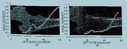
Our underlying
assumption in predicting bone fracture is that rigidity measured mechanically
(by the slope of the linear portion of the load-deflection curve) is
related to the failure load of the bone. If this assumption is true,
we can then predict the failure load of a bone by calculating rigidity
from the modulus of the bone tissue and cross-sectional moment of inertia
measured using non-invasive imaging methods such as quantitative computed
tomography (QCT), dual energy x-ray absorptiometry (DXA) and magnetic
resonance imaging (MRI). To test this assumption more rigorously, we
performed a series of experiments to examine whether rigidity was related
to the yield and ultimate loads of bones with simulated osteolytic defects,
then applied our techniques in the evaluation of benign bone tumors
in children.

