HOJ HOME | Chiefs Reports | Osgood Day | Cartilage Regeneration and Repair, Where Are We?
A Harvard Orthopaedic Presence in China
|
Scientific Articles | Alumni

click here to view full page ad

The Biomechanics of Acromioclavicular Instability

Ashwin Deshmukh, MD • David Wilson, DPhil • Jeffrey Zilberfarb, MD • Gary Perlmutter, MD

Orthopaedic Biomechanics Laboratory • Beth Israel Deaconess Medical Center

          Although the acromioclavicular joint is accessible and prominent, it is easily overlooked. Injury to this diminutive joint is often given low priority in polytrauma patients and contact athletics alike, perhaps because the long-term morbidity and functional loss from most injuries is small and because operative intervention is rarely indicated. On the other hand, as any busy clinician can attest, acromioclavicular joint pathology is a common source of shoulder pain. On occasion, particularly in high-demand sports or occupations, the pain can be disabling. It remains uncertain why certain acromioclavicular joint injuries are more symptomatic than others, and how best to treat these patients. The anatomy and biomechanics of the normal and reconstructed acromioclavicular joint are incompletely understood.1-3 Recent work in the Orthopaedic Biomechanics Laboratory (OBL) at Beth Israel-Deaconess Medical Center has investigated the acromioclavicular joint in two areas: 1) The relative stability provided by various techniques for the fixation of complete acromioclavicular disruptions, and 2) The potential for instability after subacromial decompression due to injury to the acromioclavicular ligaments.

Background

Anatomy of Acromioclavicular Stability

           The acromioclavicular joint is long and flat, with little inherent stability. The articular surfaces change from hyaline cartilage to fibrocartilage in early adulthood and there is often an intraarticular disc or meniscus. Although the point has been debated in the past, most authors now agree that very little motion occurs through the acromioclavicular joint.4

          The relatively thin capsule is reinforced by the superior, inferior, anterior, and posterior acromioclavicular ligaments. The most important stabilizers of the acromioclavicular joint are the extra-capsular ligaments that run between the coracoid process of the scapula and the clavicle.4 Two distinct ligaments exist, each named for its shape. The conoid ligament runs from the posteromedial base of the coracoid process to the underside of the junction of the middle and lateral thirds of the clavicle. The trapezoid ligament arises from the coracoid process, anterior and lateral to the origin of the conoid ligament, and attaches to the undersurface of the lateral clavicle. The deltoid and trapezius muscles span the acromioclavicular joint and provide a dynamic source of stability.

Fig. 1A
Fig. 1B

Figure 1: Type III acromioclavicular injuries are defined by complete rupture of both the acromioclavicular and the coracoclavicular ligaments.

A: There is an associated deformity, particularly in thin patients.
B: Nonoperative treatment is usually effective, but some patients develop shoulder dysfunction requiring operative treament. The goal of operative treatment is to restore the stabilizing link that the coracoclavicular ligaments provide between the clavicle and the upper extremity.

Injuries of the Acromioclavicular Joint
           Acromioclavicular injuries are usually the result of a fall on the point of the shoulder. Injuries to the acromioclavicular joint have been classified according to the extent of ligament damage, but the separate types are defined radiographically.4 A type I injury involves partial tearing of the supporting ligaments and the joint remains well-aligned; type II represents a complete tear of the acromioclavicular ligaments with partial tearing of the coracoclavicular ligaments; and type III injuries represent complete acromioclavicular dislocation (a so-called shoulder separation) with rupture of both the acromioclavicular and coracoclavicular ligaments. (Figure 1)

          Radiographs of type II and III injuries demonstrate malalignment of the acromioclavicular joint. Radiographic distinction of type II and III injuries may require the use of stress radiographs although these are rarely utilized in clinical practice. Radiographs taken of both shoulders with four kilogram weights hanging from each wrist will demonstrate an increased distance between the coracoid process and the clavicle if the coracoclavicular ligaments are completely torn.

          Types IV through VI were added by Rockwood to describe extremely displaced variants that he felt were associated with substantial muscular injury and were far more likely to require surgical treatment.4

          Treatment of type I and II injuries is non-operative, and types IV through VI are usually treated operatively, but the best initial treatment of type III injuries is disputed. While the majority of patients with type III acromioclavicular joint injuries appear to do well with non-operative treatment, a small percentage of patients initially treated non-operatively require subsequent operation because of pain and shoulder dysfunction. A number of reports suggest that certain patients, including overhead laborers, overhead athletes, and polytrauma patients tend to do poorly with non-operative treatment.


NEXT PAGE | TOP OF PAGE | HOJ HOME
Chiefs Reports | Osgood Day | Cartilage Regeneration and Repair, Where Are We?
A Harvard Orthopaedic Presence in China
|
Scientific Articles | Alumni


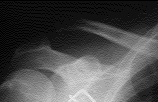
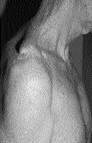
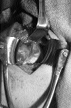
The Biomechanics of Reconstructive Techniques for Acromioclavicular Instability

          Over 150 different surgical procedures have been described for the treatment of acromioclavicular instability, and there is no widely agreed upon standard.4 Most represent some form of internal fixation between either the acromion or the coracoid process and the clavicle with or without repair or reinforcement of the ligaments using adjacent tissue. (Figure 2) Distal clavicle excision and dynamic muscle transfers have also been suggested, but are rarely used for type III injuries. The relative stability provided by various repair techniques is important, but has not been well studied.

          Investigations in the Orthopaedic Biomechanics laboratory have developed an Acromioclavicular Loading System (ALS) to study the biomechanics of the acromioclavicular joint.2 This system represents a modification of an earlier German design.3 Whereas the earlier design loaded the distal clavicle in only the superior direction, the OBL ALS allows anterior and posterior loads to be applied as well. A further advantage of our system is that accurate tracking of acromioclavicular displacements can be made using an infrared optical measurement system.

          We used the OBL ALS to compare the mechanical performance of six different surgical treatments for acromioclavicular joint instability. These included the Weaver-Dunn technique5, coracoclavicular cerclage, and four different reconstructions using suture anchors. (Figure 3) We used fresh frozen cadaveric shoulders with the scapula held rigidly in a clamp in an anatomical position. A type III acromioclavicular joint injury was simulated by cutting the acromioclavicular and coracoclavicular ligaments. The proximal end of the clavicle was held with a universal joint simulating the sterno-clavicular articulation. A wire and pulley system was used to apply anterior, posterior, and superior forces to the distal clavicle using a hydraulic materials-testing machine. Clusters of retroreflective markers were fixed to the specimen and relative motion of the clavicle and acromion was determined by measuring marker motion with the infrared optical measurement system. A digitization protocol designed by one of us (D.R.W.) was used to determine the articular surface geometry of the acromioclavicular joint. Marker displacement and joint geometry data were combined to predict the displacement between the acromial articular surface and that of the distal clavicle. This displacement in response to a known load is used as a measure of joint stability.

Figure 2: Numerous operative techniques have been described, but the mechanical stability of the various options has not been studied. This radiograph depicts failure of an acromioclavicular screw.

          Tests were performed to determine the stability of each reconstruction as well as its pullout strength. Preliminary results suggest that transfer of the coracoacromial ligament to the distal clavicle without adjunctive fixation of the clavicle to the coracoid (the so-called Weaver-Dunn technique) is inferior to the coracoclavicular suture cerclage and suture anchor techniques; however, none of the reconstructive techniques tested was able to reproduce the stability provided by the intact ligaments. The Weaver-Dunn reconstruction allowed over five times more displacement than the native ligaments, while the suture anchors and suture cerclage allowed two to three times more displacement. Similar results were found in the pullout test. Based upon these results, some form of internal fixation of the clavicle to the coracoid process seems worthwhile to protect healing ligaments and ligament reinforcements.

Fig. 3A
Fig. 3B
Fig. 3C
Fig. 3D

Figure 3: The standard Weaver-Dunn technique consists of transfer of the coracoacromial ligament to the clavicle without additional fixation of the coracoclavicular interval. Newer techniques add fixation between the coracoid and clavicle using suture anchors.

A: Operative exposure of the ruptured coracoclavicular ligaments
B: The coracoacromial ligament is isolated for transfer.
C: Suture anchors are used to secure the coracoid to the clavicle. The coracoacromial ligament is transferred to the clavicle.
D: Post-operative radiograph demonstrating restoration of stable, anatomic alignment. The distal clavicle was excised.

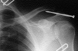
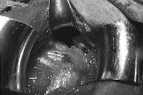
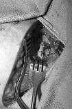
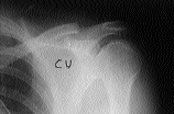
Instability of the Acromioclavicular Joint After Subacromial Decompression

          Anatomic and clinical observations suggest that open and arthroscopic techniques of subacromial decompression might result in destabilization of the acromioclavicular joint.6 Prior research in this area indicated that localized injury to the acromioclavicular ligaments or the articular surface of the acromioclavicular joint tends to destabilize the joint more in the anteroposterior than in the inferosuperior axis. Studies currently underway are measuring acromioclavicular joint stability before and after simulated standard open subacromial decompressions on several cadavers using the system described above. Our preliminary results show increased laxity of the acromioclavicular joint in both the anteroposterior and inferosuperior axes after subacromial decompression.


NEXT PAGE | TOP OF PAGE | HOJ HOME
Chiefs Reports | Osgood Day | Cartilage Regeneration and Repair, Where Are We?
A Harvard Orthopaedic Presence in China
|
Scientific Articles | Alumni


Conclusions

We have developed useful means for measuring stability of the acromioclavicular joint. Hopefully, these data and data from future studies using the OBL ALS will help limit problems associated with acromioclavicular instability.

Acknowledgements

This study was partly supported by grants from Mitek, Innovasive, Linvatec, and Arthrex.

Ashwin Deshmukh, MD is a Resident in the Harvard Combined Orthopaedic Residency

David Wilson, DPhil is a Senior Research Associate at the Orthopaedic Biomechanics Laboratory, Beth Israel Deaconess Medical Center and Instructor of Orthopaedic Surgery at Harvard Medical School

Jeffrey Zilberfarb, MD is an Attending Surgeon at Beth Israel Deaconess Medical Center, and Clinical Instructor of Orthopaedic Surgery at Harvard Medical School

Gary Perlmutter, MD is an Attending Surgeon at Massachusetts General Hospital an Clinical Instructor of Orthopaedic Surgery at Harvard Medical School

Address Correspondence to:
David Wilson, DPhil; Orthopaedic Biomechanics Laboratory; Beth Israel Deaconess Medical Center; 330 Brookline Ave. RN 115; Boston, MA 02215
e-mail: drw@obl.caregroup.harvard.edu

References
1. Bargren J, Erlanger S, Dick H. Biomechanics and comparison of two operative methods of treatment of complete acromioclavicular separation. Clin Orthop 1978;130:267-272.
2. Moses J, Wilson D, Zilberfarb J, Hayes W. A biomechanical comparison of acromioclavicular joint reconstruction techniques. The 44th Annual Meeting of the Orthopaedic Research Society. New Orleans, Louisiana, 1998.
3. Kiefer H, Claes I, Burri C, Holzwarth J. The stabilizing effect of various implants on the torn acromioclavicular joint. A biomechanical study. Arch Orthop Trauma Surg 1986;106:42-46.
4. Rockwood C, Williams G, Young D. Injuries to the acromioclavicular joint. In: Rockwood CA GD, Bucholz RW, Heckman JD, ed. Rockwood and GreenŐs Fractures in Adults. Fourth ed. Philadelphia: Lippincott-Raven Publishers, 1996:1341-1412. vol 2).
5. Weaver J, Dunn H. Treatment of acromioclavicular injuires, especially complete acromioclavicular separation. J Bone Joint Surg 1972;54A:1187-1194.
6. Kuster M, Hales P, Davis S. The effects of arthroscopic acromioplasty on the acromioclavicular joint. J Should Elbow Surg 1998;7:140-143.

TOP OF PAGE | HOJ HOME
Chiefs Reports | Osgood Day | Cartilage Regeneration and Repair, Where Are We?
A Harvard Orthopaedic Presence in China
|
Scientific Articles | Alumni