Osgood/Thesis Day | Robert Bailey Osgood Lecture | Senior Residents Thesis Presentations
Back to HOJ HOME

Robert Bailey Osgood Lecture

The Critical Role of Articular Cartilage Aging in the Development
of Osteoarthritis and in the Results of Efforts to Restore
Cartilaginous Articular Surfaces


Joseph A. Buckwalter, MD
Department of Orthopaedics • University of Iowa


          The rapid increase in the mean age of our population, and the even more striking increase in the portion of our population over age 60, is profoundly changing our health care system. A wide variety of impairments and diseases have a close relationship to aging and the prevalence and impact of these impairments and diseases is increasing as the population ages. Concern over the effects of aging on health has led physicians, scientists and the public to direct their attention toward improving understanding of age-related changes in tissues and organ systems and the role of these changes in disease and disability. Dramatic progress has been made in defining alterations in the brain, bone and skeletal muscle that occur with age and in developing approaches to decrease the adverse effects of these changes. Far less attention has been paid to the age related changes in articular cartilage. Yet no disease is more closely correlated with advancing age than osteoarthritis (the syndrome characterized by joint pain and loss of joint function due to joint degeneration), and no disease causes more impairment of mobility than osteoarthritis. Furthermore, age changes in articular cartilage have important implications for efforts to restore cartilaginous articular surfaces for middle age and older patients.

Age and the Prevalence of Osteoarthritis
          The close relationship between age and the prevalence of osteoarthritis (Figures 1 & 2) has led to the widespread concept that osteoarthritis is an inevitable result of growing older; that is, with time and use joints wear out. This concept leads to the conclusions that preventing the development or progression of osteoarthritis is not possible and that the best approaches to management of the disease are symptomatic treatments for patients with mild or moderate disease and joint replacement for patients with severe disease. An alternative view is that the joint degeneration responsible for osteoarthritis is distinct from articular cartilage aging. The implication of this point of view is that the potential exists to develop strategies that will decrease the risk or slow the progression of osteoarthritis. Determining which of these views is correct requires understanding of articular cartilage aging, and its distinction from the joint degeneration responsible for osteoarthritis.

Figure 1. The overall prevalence rate for osteoarthritis diagnosed by examination increases with age and is about twice as high in women (This histogram was developed from information published in Praemer AP, Furner S, Rice DP: Musculoskeletal Conditions in the United States. 1992, Park Ridge, Illinois: American Academy of Orthopaedic Surgeons.)

Osgood/Thesis Day | Senior Residents Thesis Presentations
NEXT PAGE | TOP OF PAGE | HOJ HOME


Figure 2. The prevalence rates for radiographic changes of osteoarthritis in the hand (2A), foot (2B), knee (2C) and hip (2D) increase with age. Radiographic changes occur most commonly in the hand and with decreasing frequency in the foot, knee and hip. Women have higher prevalence rates for radiographic changes in the hand, foot and knee in almost all age groups. (These histograms were developed from information published in Praemer AP, Furner S, Rice DP: Musculoskeletal Conditions in the United States. 1992, Park Ridge, Illinois: American Academy of Orthopaedic Surgeons.)
Articular Cartilage Aging
          Articular cartilage undergoes significant structural, matrix, cellular and mechanical changes with age. Autopsy studies have documented increasing prevalence of superficial and even deep fibrillation of the articular surfaces of all synovial joints with age.(Figure 3) This condition is more common in some joints than others, but appears to be a universal age-related process. Although age-related cartilage fibrillation may progress, the vast majority of patients with this condition do not develop joint pain or dysfunction. Thus, these age-related structural changes are distinct from those seen in the joint degeneration responsible for osteoarthritis. Alterations in aggregating proteoglycans (molecules referred to as aggrecans that consist of central protein cores with multiple covalently bound chondroitin and keratan sulfate chains that give articular cartilage its stiffness to compression, resilience and durability) are among the most striking articular cartilage matrix changes with age. The size of proteoglycan aggregates (molecules formed by the non-covalent association of multiple aggrecans with a hyaluronan filament) decreases significantly with age (Figure 4), because the aggrecan molecules become shorter (Figures 4 & 5), as do their chondroitin sulfate chains, and because the mean number of aggrecans in each aggregate decreases. These age-related changes might be due to degradation of the proteoglycans in the matrix, alterations in proteoglycan synthesis, or both.

           Evidence from studies of bovine chondrocytes in vitro suggests that there is an age-related change in aggrecan synthesis that leads to the production of shorter protein cores and chondroitin sulfate chains. Likewise, in vitro experiments show that aggregates assembled in vitro in cultures of older chondrocytes are smaller and more irregular than those assembled in cultures of younger chondrocytes. Some of these changes in aggregates may be caused by age-related alterations in link proteins (the small proteins that stabilize the association between aggrecans and hyaluronan). Other age related changes in the molecular composition and structure of the articular cartilage matrix include increased collagen cross-linking and decreased water concentration.

Figure 3. Prevalence of humeral and patellar articular surface fibrillation and degeneration determined at autopsy. (This histogram was developed from data originally published by Collins and Meachim (1961) and reported by Freeman in Freeman MAR: Adult Articular Cartilage. 2nd ed. 1979, Tunbridge Wells: Pitman Medical Publishing. pp 560.)

Figure 5. Histogram showing the decrease in human proteoglycan aggrecan length with age.

          Recent studies of rat articular cartilage chondrocytes revealed an age related decline in the anabolic response of the cells to insulin dependent growth factor I (IGF-I).(Figure 6) IGF-I appears to be one of the most important anabolic factors in articular cartilage and thus presumably has a critical role in maintaining the chondrocyte synthetic activity that preserves articular cartilage. The age related decline in the anabolic response of articular cartilage chondrocytes to IGF-I may be associated with increased expression of insulin growth factor binding proteins. An age related increase in the production of these binding proteins could decrease the ability of the chondrocytes to maintain or repair the articular cartilage matrix. In addition to the structural, molecular and cellular changes in articular cartilage with age, the mechanical properties of the tissue, including tensile stiffness and strength, deteriorate with age.
back
Figure 4. Electron micrographs of bovine articular cartilage proteoglycan aggregates. Aggregates consist of central hyaluronan filaments with multiple attached aggrecans. A. Proteoglycan aggregate from a cell culture of two to three month old calf chondrocytes. B. Proteoglycan aggregate from a cell culture of two to three year old steer chondrocytes. C. Proteoglycan aggregate from articular cartilage of a two to three month old calf. D. Proteoglycan aggregate from the articular cartilage of a two to three year old steer. Notice that aggrecans from calf cell cultures (A) and from calf articular cartilage (C) are longer and vary less in length than steer aggrecans (B & D) and that aggregates from calf cell culture (A) and from calf articular cartilage (C) are larger than aggregates from the corresponding steer chondrocyte culture (B) and articular cartilage (D).

back

Figure 6. Graph showing that with increasing age rat tibial articular cartilage chondrocytes incorporate less sulfate and that their response to the anabolic cytokine IGF-I declines with increasing age.


Osgood/Thesis Day | Senior Residents Thesis Presentations
NEXT PAGE | TOP OF PAGE | HOJ HOME


Osteoarthritis
          In contrast to the aging changes in articular cartilage, osteoarthritis is a clinical syndrome due to the degeneration of synovial joints. One of the first events in articular cartilage degeneration is disruption or alteration of the molecular structure and composition of the matrix. Some of the early matrix changes in articular cartilage degeneration include loss of proteoglycans and an increase in water concentration. The tissue damage stimulates a chondrocytic synthetic and proliferative response that may maintain or even restore the articular cartilage. This chondrocytic response may continue for years; however, in progressive joint degeneration, eventually the chondrocytic anabolic response declines and the imbalance between chondrocyte synthetic activity and degradative activity leads to progressive thinning and loss of articular cartilage. Even in the early stages of the joint degeneration the stiffness of the articular cartilage declines and its permeability increases. These alterations in material properties may further accelerate the progression of the disease. The response of the synovial joint to joint degeneration can in some instances restore a form of cartilaginous surface. It is not clear how frequently this occurs, but well documented studies of small groups of patients confirm that even in individuals with complete loss of articular cartilage the potential exists for spontaneous restoration of a cartilaginous surface that may function effectively for years. Thus far the characteristics of patients in whom this response occurs have not been defined, but this phenomenon deserves further study.
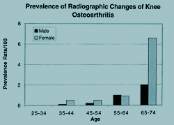
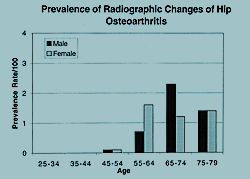
          If articular cartilage aging and osteoarthritis are distinct processes (Table 1), how can the striking increase in the prevalence of the joint degeneration responsible for osteoarthritis with age be explained? The answer appears to be that the structural, molecular, cellular and mechanical changes that occur in articular cartilage with age increase the vulnerability of the tissue to degeneration. Furthermore, the evidence that articular 
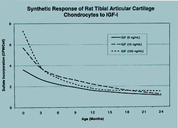
cartilage chondrocytes are less responsive to anabolic cytokines with increasing age suggests that older articular cartilage is less able to repair and restore itself. Thus, the aging of articular cartilage does not cause osteoarthritis, but the aging changes in articular cartilage increase the risk of joint degeneration, and decrease the ability of joint tissues to prevent progression once degeneration begins.

    Aging
Osteoarthritis

Structural Localized fibrillation
     
     
Fibrillation and fragmentation extending to subchondral bone
Loss of tissue (decreased cartilage thickness and complete loss of cartilage in some regions)
Formation of fibrocartilagenous repair tissue

Matrix Decreased water concentration
  Loss of large proteoglycan aggregates (decreased aggregate stability?)
  Increased decorin concentration
  Accumulation of degraded molecules (aggrecan and link protein fragments)
  Increased collagen cross-linking
  Increased collagen fibril diameter and variability in fibril diameter
  Decreased tensile strength and stiffness in superficial layers
Initial increase in water content
Disruption of collagenous macromolecular organization
Progressive degradation and loss of proteoglycans and hyaluronan
Progressive degradation and loss of collagens
Increased fibronectin concentration
Increased permeability and loss of tensile and compressive stiffness and strength

Cells Decreased chondrocyte density with skeletal growth
  Alteration in synthetic activity (smaller more variable aggregcans)
  Decreased anabolic response to growth factors (IGF-I)
  Decreased synthetic activity
Initial increase in synthetic and proliferative activity
Loss of chondrocytes
Eventual decreased synthetic activity
Increased degradative enzyme activity
Appearance of fibroblast like cells in regions of fibrocartilagenous repair tissue

Effects of Aging on the Results of Methods of Restoring Articular Cartilage
          Understanding of the aging of articular cartilage and its relationship to osteoarthritis has significant implications for strategies intended to restore cartilaginous articular surfaces. Currently, these strategies can be separated into two groups: those performed with the intention of stimulating articular cartilage repair or regeneration, and those that include transplantation of osteochondral autografts or allografts. The outcomes of both strategies may be significantly influenced by the changes that occur in articular cartilage with aging.

          Current clinical procedures intended to promote articular cartilage repair and regeneration include penetration of subchondral bone, altering joint loading by osteotomies or joint distraction and use of perichondrial and periosteal transplants. Experimental methods promoting formation of cartilaginous articular surfaces include use of artificial matrices, chondrogenesis factors and cell transplants. Penetration of subchondral bone by a variety of methods including abrasion, drilling and penetration with a sharp awl or pick (a procedure referred to as microfracture) stimulates formation of a fibrin clot followed by invasion of the fibrin clot by undifferentiated cells that proliferate, differentiate into chondrocyte like cells and synthesize a matrix that contains high concentrations of many of the molecules found in normal articular cartilage including type II collagen and aggregating proteoglycans. The chondral repair tissue that forms following these procedures is less stiff and more permeable than normal articular cartilage. This may make the tissue more vulnerable to degeneration although some patients do develop excellent symptomatic relief. Because of the limitations of the chondral tissue formed following penetration of subchondral bone, some surgeons perform autograft transplants of perichondrium and periosteum with the intent of generating a new articular surface. The available evidence suggests that both penetration of subchondral bone and periosteal and perichondrial transplants are less clinically successful in middle aged and older people than in adolescents and young adults. There is less experience with autograft cell transplantation, use of artificial matrices and chondrogenesis factors, but given the reliance of these procedures on host cells it is reasonable to expect that they will less successful in older individuals than in young adults. Use of fetal cell allograft transplants offers the potential of overcoming some of the limitations of relying on host cells from older patients, but the efficacy of this approach has not yet been demonstrated.

          A variety of methods of performing osteochondral allografts and autografts have been developed to replace damaged or degenerated articular surfaces. The experience of Allan Gross in Toronto and others with osteochondral allografts suggests that when these transplants are performed in young adults to replace focal defects in otherwise normal joints they can successfully provide pain relief and improve joint function for more than 60% of the patients for as long as 20 years. A number of surgeons have also been transplanting osteochondral autografts from a region of normal articular to regions of damaged articular cartilage. Although the effects of patient age on the results of both allografts and autografts have not been extensively studied, the available information suggests that both procedures are less successful in middle-aged and older people.

Conclusions
          Articular cartilage undergoes age-related changes that increase the risk of the joint degeneration that causes the clinical syndrome of osteoarthritis. In addition, these changes adversely affect the outcomes of attempts to repair or regenerate articular cartilage. Perhaps the most important of these age changes involve alterations in chondrocyte synthesis of proteoglycans and in the responsiveness of chondrocytes to anabolic factors. Is it possible to slow, or temporarily reverse at least some of the age-related changes in articular cartilage that increase the probability of joint degeneration? Once degenerative changes have developed, can they be stabilized or reversed? Will it be possible to develop methods that predictably produce functional, durable cartilagenous articular surfaces for middle age and older people with joint injuries and joint degeneration? Further study will definitively answer these questions. However, the investigation of articular cartilage aging is only beginning and the observations developed thus far strongly suggest that better understanding of the aging changes in articular cartilage and how these changes influence the ability of the tissue to maintain and regenerate itself will lead to improved methods of preserving and restoring articular surfaces for middle-aged and older individuals.

Address correspondence to:
Joseph A. Buckwalter;
01013 Pappajohn Pavilion; Department of Orthopaedics;
University of Iowa College of Medicine; Iowa City, IA 52242
e-mail: joseph-buckwalter@uiowa.edu

Osgood/Thesis Day | Senior Residents Thesis Presentations
TOP OF PAGE | HOJ HOME