
Research
28th Annual Meeting of The Orthopaedic Trauma Association
September 6-8, 2012, Chicago, IL
Accuracy and Reliability of Bohler’s Angle Measurements With Oblique Lateral Radiographs Taken in the Trauma Setting
R. James Toussaint, M.D., Ida L. Gitajn, M.D., John Y. Kwon, M.D.
Massachusetts General Hospital, Boston, MA, USA
Purpose
First described in 1931, Bohler’s angle is used to determine the amount of posterior facet displacement and severity of injury in calcaneus fractures. It is used to guide management or the need for additional imaging. Lateral images used to obtain Bohler’s angle in the trauma setting are often oblique due to difficulties in positioning of the traumatized extremity or limitations from splint materials. Inaccurate Bohler’s angles in this setting can lead to under/overtreatment of patients. The purpose of this study is to assess the accuracy and reliability of measuring Bohler’s angle on oblique lateral imaging and to determine how improper imaging influences measurement.
Methods
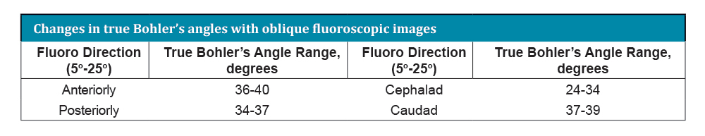
A cadaver specimen was imaged using a large C-arm to obtain multiple fluoroscopic images. First, a perfect lateral was obtained. Next, a series of oblique images was taken with the beam directed anteriorly, posteriorly, cephalad, and caudad. The images were taken in 5° increments from 5° to 25° in each direction. Orthopaedic staff and residents were asked to measure the observed Bohler’s angles. To define the true Bohler’s angles, metallic markers were then placed on the anterior calcaneal process, the superior most portion of the posterior facet, and the superior posterior tuberosity of the same cadaver calcaneus. The same series of images were repeated to measure true Bohler’s angles using the marked specimen. The senior author then measured the true Bohler’s angles from the marked specimen.
Results
41 orthopaedic staff and residents participated in the study. The mean values for the observed Bohler’s angles were significantly different (P <0.05) from the true Bohler’s angles for all series of images except a posteriorly directed x-ray beam at 20° from the horizontal (P = 0.43). The mean value for observed Bohler’s angles deviated further from the true Bohler’s angles with increasing image obliquity for all series except the posteriorly directed x-ray beam. The true Bohler’s angle on a perfect lateral image was 35°. The true Bohler’s angle was found to vary based on the obliquity of the fluoroscopic image (see table below).
Conclusion
The study findings reveal that orthopaedic staff and resident physicians’ ability to accurately measure Bohler’s angle significantly decreases with increasing obliquity of lateral radiographs. The true Bohler’s angle also varies with image obliquity. Understanding these changes with oblique lateral radiographs taken in the trauma setting should decrease reliance on only Bohler’s angle to determine management and need for additional imaging.

- In This Issue
- Senior Thesis Day
- Abstracts
- Research Reviews
- Citations