HOJ HOME | Chiefs Reports | Osgood Day | Scientific Articles | Alumni Association
Click here to visit our web site

A Look at the Past and a Peek into the Future

John E. Hall, MD

Children's Hospital Department of Orthopaedic Surgery


As we enter this centennial year, it seems to be an appropriate time to look backwards as well as forward. There is good reason to believe that the early part of this century will see the same rapid change in orthopedic surgery as in all other areas of endeavor. Since this year will be my forty-second year of practice, it is easier for me to look back than to try to predict the future. 

Similar to other medical specialties, Pediatric orthopaedic surgery has seen astounding changes since I began my practice in 1958 at the Hospital for Sick Children in Toronto with Bob Salter, and continued it at Children's Hospital in Boston since that time.

Dr. Hall is Professor and Chairman Emeritus, Children's Hospital Department of Orthopaedic Surgery.

Please address correspondence to:
John E. Hall, MD
Department of Orthopaedic Surgery
The Children's Hospital
300 Longwood Ave.
Boston, MA 02115 hall_ j@a1.tch.harvard.edu
 

Much of my residency and early practice was devoted to the management of the sequelae of poliomyelitis. It is difficult to describe the anxiety and precautions taken in communities in the years of polio epidemics, and the number of children left with permanent disabilities. The disappearance of this scourge has to be one of the defining events of this period. Much the same can be said about tuberculosis as evidenced by the absence of the dedicated sanitoria, which were so prevalent at mid-century. Whether either of these diseases could reappear will depend on compliance with immunization programs on the one hand and development of resistant strains and poor public health measures on the other.

One of my major interests has been the management of spinal deformities. The length and character of hospital stays following pedi-atric spine surgery has changed from one yearto a week or less. Despite common opinion, this has little to do with the emphasis on cost-containment in today's competitive hospital environment. The greatest stimulus to shortened hospital stays in Pediatric Orthopedics has been the desire of parents to have their children at home as soon as feasible, and the development of techniques that have made this desire a reality.
Figure 1. Recumbent turnbuckle plasters for the treatment of spinal deformity.

From recumbent turnbuckle (Fig. 1) plasters to ambulatory Risser casts (28) to stable internal fixation, the lot of the child with a severe spinal deformity has been made easier. After learning the use of Cobb turnbuckle jackets at the Royal National Orthopaedic Hospital in London, England, with Mr. J.I.P. James (19) , I returned to Toronto where patients were suspended from the ceiling in a fish net to have their plasters applied (Fig. 2)( 21) . You can easily see how ready I was for Paul Harrington when he introduced his internal rods in the late 1950's. Visits to John Moe in Minneapolis (27) , Wally Blount in Milwaukee (3) and Joe Risser in Los Angeles (28) helped further my education in education in the management of scoliosis. With money from the Rehab Symposium in Toronto I was able to invite Arthur Hodgson (16,17) from Hong Kong (who pioneered the anterior approach to the spine for tuberculosis, Paul Harrington from Texas whose rods were the first effective internal fixation system (15) and Alan Dwyer from Sydney, Australia who introduced anterior instrumentation (9) to teach me these methods. A session in Paris with my friends Yves Cotrel and Jean Dubousset in the early 1980s opened new vistas with their double rod rotating system (7) . They laid the groundwork for the amazing proliferation of the currently used spinal instrumentation systems. Extensive surgery, however, on a back deformed by "idiopathic scoliosis", has been the result of our failure to find the root cause of and to prevent the deformity from occurring in the first place. Perhaps genetic engineering will provide the answer to this challenging problem in the coming century.
Figure 2. Patient suspended in fish net prior to plaster application for spinal deformity.

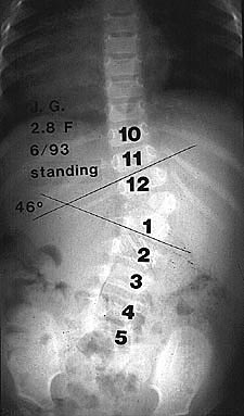
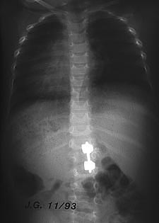
The Scoliosis Research Society, first formed in 1966 with 35 members, has grown now to over 600 fellows and others devoted to spinal deformity problems. It is a powerful force in the dissemination of information and the encouragement of research in this field. Affiliations with similar societies in many other countries have helped to foster an international flow of information.

Improvements in anesthesia techniques have allowed much more extensive surgery in all fields. In spinal surgery anterior and posterior spinal procedures can be performed under the same anesthetic, often in children who were formerly considered unsuitable candidates for either operation. They have allowed my current favored method of treating unbalanced spines in very young children with progressive hemivertebral anomalies with a hemivertebral resection with the front and back open simultaneously. The infant instrumentation system used in this technique was designed by Eduardo Luque (23) has made it easy to obtain and maintain corrections as seen in Figures 3a and b.

Spinal bracing has evolved amid contoversy concerning its effectiveness. What has clouded this issue has been confusion concerning the difference between the bad results of bracing and the results of bad bracing.

Figure 3. Preoperative (a) and postoperative (b) radiographs of surgical treatment of a hemivertebral anomaly using infant Luque instrumentation.

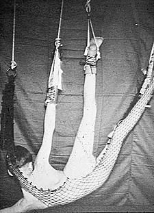
The Boston System of bracing was designed with Bill Miller (13) , a gifted orthotist, and has led to the almost complete disappearance of the neck ring in current bracing.

The treatment of childhood amputees, particularly those with congenital deformities, has evolved from wooden legs to complex well-engineered lightweight prostheses (31) . Power-assisted upper extremity and swing-and-stance- phase lower extremity models have improved performance, although at greatly increased cost. Attempts have even been made to construct powered orthoses for paraplegics to give them some form of independent ambulation. Powered wheelchairs and mobility devices have been made available to even the most severely disabled and computerized controls have even provided speech. Continued development along these lines can be expected. Experimental work in animals has tried to solve the problem of repair of central nerve elements such as the spinal cord. Perhaps this will become possible in this century.

The tragedy of the severe skeletal and other abnormalities caused by the use of thalidomide in pregnant women (Fig. 4) has alerted the medical profession to preventable causes of congenital malformations (30) . When the problem surfaced in Canada in the early 1960's, I was asked to join a task force to advise the Canadian Government on how to handle it (14) . Our group visited Germany and other countries to learn more about how they were coping with their large numbers of deformed children. Further work in the hereditary and developmental factors may lead us to methods of preventing at least some of these disorders.

Early detection of developmental dysplasia of the hip by screening examinations and ultrasound have led to early splinting and better control of this problem (12) . Surgical techniques for patients who have failed early detection and treatment have improved the salvage of these hips, even in early adult life, hopefully preventing or delaying the onset of osteoarthritis. Bob Salter's innominate osteotmy (29) has focused attention on the acetabulum as a major part of the problem. (Fig. 5)

Figure 4. Birth abnormalities secondary to thalidomide exposure.
During my training period in England we removed many broken Judet prostheses (20,26) and returned to McMurray's osteotomy (25,4) to treat osteoarthritis of the hip. John Charnley's (8) introduction of cemented total joint replacement led to the explosion of interest and technical development which has been such a boon to sufferers from joint deterioration. Perhaps even the current controversy over cemented versus cementless component designs will be settled in the coming decades.
Figure 5. Schematic diagram depicting the correction obtained by the Salter innominate osteotomy for hip dysplasia.

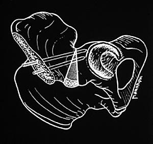
The management of clubfeet has developed from manipulative correction and retention with various forms of strapping splints and casts to a clearer idea of the role and timing of surgical correction. Although Codivilla (6) has described a medial release in the early part of the century, it was Vince Tureo (33) who popularized the method and made it more effective by using a K-wire to hold the reduction of the talo-navicular joint. Ultrasound has made possible the detection of these and other anomalies in the uterus and some fetal surgery has already been used in potentially lethal conditions. This field may extend to some orthopedic anomalies in the coming century.

Malignant tumors were so uniformly fatal in my early career, that survival often caused one to question the original diagnosis. The development of chemotherapy has prolonged survival and allowed limb salvage techniques (11). Surely the next century will see an expansion of our knowledge in this area and even lead to the eventual eradication of many malignancies.

One of the most dramatic developments in orthopaedic surgery has been the introduction and perfection of arthroscopic surgery. It has come from Watanabee's (34) incandescent bulb on the end of a scope in the knee for diagnostic purposes to the sophisticated video assisted instruments in use today in almost all joints. Improved surgical techniques have allowed shortened hospital stays and even outpatient procedures, improving rehabilitation and outcomes.

Fracture care in children has always included a large reliance on closed manipulation and some form of cast retention. There has been a better understanding of the role of surgical correction in specific fractures such as the neck of the femur, the elbow region and intra-articular injuries. Intramedullary fixation techniques formerly limited to severe multiple injuries and older children have been extended to include femoral fractures in much younger children (22) . Whether these techniques will replace non-operative management in even wider indications remains to be seen.

Imaging techniques such as computerized tomography and magnetic resonance imaging have helped enormously in fracture care, as it has in spinal surgery and almost all other conditions. Further development along these lines can be expected.

Leg lengthening, once a rarely performed procedure has become more common place with the development of improved knowledge of bone healing and surgical techniques. Anderson's method of osteotomy of a long bone and gradual distraction in use in the 1940's and 1950's 1 was hampered by the necessity of prolonged immobilization in a frame. Ilizarov improved on this method by delaying the start of distraction until callus had formed and by designing a frame in which the patient could be ambulatory (18) . Unilateral frames have made femoral lengthenings more tolerable and lengthening over an intramedullary nail more stable. Its exact role should become clearer in the future

Resuscitation techniques have saved many children with multi system trauma, utilizing greater knowledge of electrolyte and volume management (32). Head injuries remain the most serious problems, and one research focus has been on understanding the permanent sequelae and prevention of them. (2,24) The use of seatbelts (incredibly enough not universal), air bags and structural improvement in automobiles have decreased mortality and morbidity and public education and enforcement in their use will save many more lives and cerebral function.

Enzyme research has led to the control of such conditions as Gaucher's disease (10) and should lead to the prevention of many more. The costs of these therapies are the major stumbling block, and it is to be hoped that as cost declines in the years to come, accessibility of these drugs to patients will increase..

The Pediatric Orthopedic Society of North America has grown from two independent societies formed in the 1970s to a large well organized group which has improved knowledge and practice in the specialty and can be expected to have an even greater impact in the future. (Fig. 6)

Intern and residency training programs have gone from haphazard arrangements to well organized and supervised education. It is to be hoped that the current problems with funding will be solved, and resident education will remain a primary objective for our teaching institutions.

Figure 6. Founding members of the Pediatric Orthopaedic Society of North America.

Teaching in Pediatric Orthopedics, traditionally a part of general medical and surgical education, and part of general orthopedic training, has evolved to a more definable specialty. Pediatric orthopedic fellowships have been established and have led to many orthopedists confining their practice to pediatric orthopaedics. Even subspecialties within the field of pediatric orthopaedics have developed and this trend can be expected to continue.

What form medical coverage will assume in the future when the present challenges have been resolved is open to speculation. One can only hope and believe that the tremen-dous development that has occurred in the past half century will blossom and expand into this century and beyond.


TOP OF PAGE | HOJ HOME
HOJ HOME | Chiefs Reports | Osgood Day | Scientific Articles | Alumni Association


References
1. Anderson W.V. Leg lengthening. J Bone Joint Surg 1952;34B:150.
2. Benowitz LI, et al. Inosine stimulates extensive collateral growth in cortico-spinal tracts
3. Blount WP, Schmidt AL, Keever ED, Leonard ET. The Milwaukee brace in the operative treatment of scoliosis. J Bone Joint Surg 1958;40:511-25.
4. Campbell, JP, Jackson J.P. Treatment of osteoarthritis of the hip by osteotomy. J Bone Joint Surg 1956;38B:468-74.
5. Cobb JR. Outline of the study of scoliosis. AAOS Instructional Lectures 1948;5:261-75.
6. Codivilla A. Sulla cura del piede equino varo congenito. Nuoro metodo di cura cruenta. Arch Ortop 1906;23:245-56.
7. Cotrel Y, Dubousset J, Guilaumat M. New universal instrumentation in spinal surgery. Clin Orthop 1988;227:10-23.
8. Charnley J. Anchorage of the femoral heart prosthesis to the shaft of the femur. Use of a cold curing cement. J Bone Joint Surg 1960;42B:28-30.
9. Dwyer AF, Newton NC, Sherwood AA. An anterior approach to scoliosis. A preliminary report. Clin Orthop 1969;62:192-202.
10. Elstem D, Itzachoki M, Mankin H. Baillieres. Clinical Hem 1997;10:793-816.
11. Gebhardt MC, Associate Editor, Simon MA, Springfield D, eds. Surgery for bone and soft tissue tumors. Philadelphia: Lippincott-Raven, 1998:375-404.
12. Graf R. New possibilities for the diagnosis of congenital hip joint dislocation by ultrasonography. J Pediatr Orthop 1983;3:354-59.
13. Hall JE, Miller M, Shuman W, Stanish W. A refined concept in the management of idiopathic scoliosis. Prosthet Orthot Int 1975;29:7-13.
14. Hall JE. Surgery of limb defects. Can Med Assoc J 1963;88-
15. Harrington P. Treatment of scoliosis: Correction and internal fixation by spinal instrumentation. J Bone Joint Surg 1962;44:592-610.
16. Hodgson AR, Stock FE. Anterior spine fusion for the treatment of tuberculosis of the spine. The operative findings and results of treatment of the first one hundred
cases. J Bone Joint Surg 1960;42A:295-310.
17. Hodgson AR. Correction of fixed spinal curves. J Bone Joint Surg 1964;47:1221-7.
18. Ilizarov GA, Deviatov AA. Operative elongation of the leg with simultaneous correction of deformities. Orthop Traumatol Progez 1969:30:3.
19. James JIP. Idiopathic scoliosis: The prognosis, diagnosis, and operative indications related to curve patterns and the age of onset. J Bone Joint Surg 1954;36:36-49.
20. Judet R, Judet J. Technique and results with the acrylic femoral head prosthesis. J Bone Joint Surg 1952;34B:173.
21. LeMesurier AB. A method of correcting the deformity in scoliosis before performing the fusion operation. J Bone Joint Surg 1941;23:521-32.
22. Ligier JN, Metaizeau JP, Prevot J, Lascombes P. Elastic stable intramedullary nailing of the femoral shaft fracture in children. J Bone Joint Surg 1988;70B:74-77.
23. Luque ER. Segmental spinal instrumentation for correction of scoliosis. Clin Orthop 1982;163:192-209.
24. Madsen JR, et al. Tacrolemas (K506) increases neuronal expression (GAP-43) and improves functional recovery after spinal cord injry in rats. Epxer. Neural 1998;154:673-683.
25. McMurray TP. Osteoarthritis of the hip joint. J Bone Joint Surg 1939; 21:1.
26. Pridie KH. The problem of the broken Judet prosthesis. J Bone Joint Surg 1955;37B:224-227.
27. Moe JH. A critical analysis of methods of fusion for scoliosis. An evaluation of two hundred and sixty-six patients. One hundred thirty patients had idiopathic, 136 had
paralytic scoliosis. J Bone Joint Surg 1958;40A:520-554.
28. Risser JC, Norquist D. A follow-up study of the treatment of scoliosis. J Bone Joint Surg 1958;40A:556-69.
29. Salter RB. Innominate osteotomy in the treatment of congenital dislocation and subluxation of the hip. J Bone Joint Surg 1961;43B:518-39.
30. The Insight Team of the Sunday Times of London. Suffer the Children: The Story of Thalidomide. Viking Press, New York, 1979.
31. Herring JA, Birch JG, editors. The Child with a Limb Deficiency. American Academy of Orthopaedic Surgeons, Rosemont, IL 1998:20.
32. Tscherne H, Regel G. Care of the polytraumatized patient. J Bone Joint Surg 1996:78B:840-52.
33. Turco VJ. Surgical correction of the resistant club foot. J Bone Joint Surg 1971;53A:477.
34. Watanabe M, Takeda S. The No. 21 arthroscope. J Japanese Orthop Assoc 1960;22:59.

TOP OF PAGE | HOJ HOME
HOJ HOME | Chiefs Reports | Osgood Day | Scientific Articles | Alumni Association