| HOJ HOME | Chiefs Reports | Osgood Day | Scientific Articles | Alumni Association |
|
| Click here to visit our web site |
| ||||||||||||||
|
I. Age-Related Incidence of Fractures Epidemiological studies
indicate that there is a biphasic pattern for appendicular fractures peaking
in the early teen years and again in later life. The majority of fractures
in childhood and adolescence are the result of antecedent trauma, whereas
the majority of fractures in the elderly occur in association with only
minimal or moderate trauma. The location of fractures is different in
these two periods of life. In the elderly, fractures of the vertebrae,
distal forearm, and proximal femur among the elderly are commonly attributed
to osteo-porosis (1) .
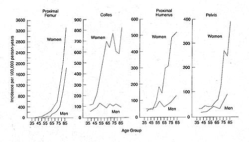
There are striking gender differences in fracture locations and rates with aging (Figure 1). There is a steep rise in Colles' fractures in women around the time of the menopause.
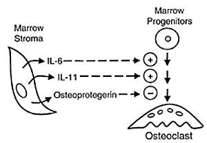
II. Age-Related Bone Loss in Women and Men All vertebrates lose bone mass with age. Cross-sectional studies indicate a progressive decline in bone mass in both women and men, with an acceleration in women after menopause (2) . Only recently have quantitative measures of bone mass become available as a practical clinical tool. Single photon absorptiometry was the first technique for measuring bone in the peripheral skeleton. This method was improved upon in the 1980's by dual-photon absorptiometry. In the last decade, methods that are based upon radioisotope sources of energy have been replaced by X-rays, with dual-energy X-ray absorptiometry (DXA) being the most common, validated tool. DXA provides measures of bone mineral content (grams) and bone area (cm squared). Bone mineral density (BMD, grams/cm squared ) is calculated from those measures and is correlated with bone strength. A patient's BMD is expressed as a Z-score, or the number of standard deviations above or below the mean value for young normal adults. The Z-score is the comparison with persons of the same age, gender, and race. Numerous studies indicate that BMD is an index of fracture risk (3) . Quantitative computed tomography (QCT), a volumetric measurement, is useful in measuring vertebral bodies with osteophytes and sclerotic facets. Quantitative ultrasound also has great potential as a screening tool. Several longitudinal studies have examined the effect of age on the rate of bone loss. In a large multi-center study of women from the age of 65 to 85 years of age, Ensrud et al. reported that the rate of loss of BMD in the femoral neck increased from 0.35% per year at 65 years of age to more than 1% per year after age 85 years (4) . Similar results have been reported using QCT and ultrasound technology (5) . III. Endocrinology of Aging Aging is accompanied by declines in the endogenous release or production of many hormones that have links to skeletal metabolism (Table I) (6) . The somatopause describes the age-related dampening of the number and magnitude of daily pulses of growth hormone (GH) secretion (7) . It has been recognized that the skeletal features of GH excess, or acromegaly, are due to overgrowth of bones, particularly of cortical architecture. In contrast, GH deficiency is associated with delayed skeletal maturation, decreased bone mass, and increased fracture incidence, compared to normal subjects. It is likely that these skeletal effects are mediated by GH's stimulation of production of the osteotropic, anabolic agent insulin-like growth factor (IGF) in the liver and directly in bones. Serum levels of IGF-I and its binding proteins, IGFBP-3, and -5, also show striking declines with age (8) . The adrenal steroid dehydroepiandrosterone (DHEA) and its sulfated form, DHEAS, are the most abundant steroids in the human circulation. The adrenopause describes the decline in circulating levels of DHEA(S). In healthy subjects, DHEAS levels peak during the early twenties and decline to 10-20% of peak levels by age 70 (9) . In a study of 102 normal women between 28 and 96 years of age, we reported that DHEAS and IGF-I levels declined with age (p<0.0001) and IGF-I was positively correlated with DHEAS (p=0.008) (10) . The hypothesis that the adrenopause may contribute to age-related bone loss through a deficiency in IGF-I is supported by the observation that administration of DHEA to the elderly resulted in increased serum IGF-I to youthful levels (11) . At menopause, there is a generalized decrease in estradiol produced by the ovary. Although the adrenal androgens can served as precursors to estrogen at that time, the net result is a decline in circulating estradiol. In men, estradiol produced in the adrenals circulates at levels somewhat lower than in women, and population studies suggest that it does not decline dramatically with age. There is controversy about the existence of the andropause, unless symptoms of hypogonadism manifest with aging. It is generally accepted, however, that androgen levels decline with aging and it has been proposed that glucuronidated derivatives of androgens may be more meaningful measures of androgen status. Those androgens show striking declines with age. In a study of 145 healthy men aged 60 to 91, 54% had low levels of androgens, half of whom also had low levels of gonadotropins (12) . Hypogonadism and impotence are common, yet independent conditions of older men (6) . Thus, andropause is a variable process when it occurs. On the other hand, most women pass through an andropause with aging, as evidenced by striking declines in serum levels of glucuronidated androgens. IV. Relationships Between Hormone Deficiencies and Bone Density Large population-based studies show strong and significant positive correlations between estradiol and bone density at the spine, forearm, and hip (12) . These associations hold for both women and men. Very low or undetectable estrogen levels are a risk factor for decreased bone density and hip fractures in women, and more recently, a similar correlation has be found in men. Bone is an androgen-dependent tissue. Hypogonadal boys and men have low bone density that can be improved with testosterone therapy (6) . There are conflicting reports on the relationship of androgen status with bone density and fracture risk in normal individuals, with positive correlations reported at some bone sites (12) . Many of these studies are confounded by other variables, such as smoking status. There are contradictory reports on the relationship between serum DHEA(S) levels and bone density. The large Rancho Bernardo study found positive correlations in women at some bone sites (12) . We recently examined a cohort of 102 normal women between 28 and 96 years old (10) . Levels of DHEAS and IGF-I were correlated significantly with bone density scores at the spine and hip sites. Ongoing trials of DHEA replacement therapy will test for improvements in bone density. V. Roles of Bone Marrow in Skeletal Aging Bone marrow cells play many roles in skeletal metabolism. They secrete cytokines that mediate communication among various cells types. The hematopoietic fraction of marrow includes osteoclast progenitors, while the stromal compartment includes cells that support osteoclast differentiation and includes precursor cells capable of osteoblast differentiation. We proposed that skeletal
aging is a consequence of aging of the bone marrow. With age, marrow stromal
cells may be less able to differentiate into osteoblasts and more capable
of supporting differentiation of osteoclasts (Figure 2). These hypotheses
were tested with human marrow that was discarded during total hip arthroplasty.
We discovered that in vitro osteoclastogenesis increases with age (13)
. Various cytokines increase osteoclast differentiation, especially in
animal marrow cultures, but little is known about mechanisms responsible
for increased osteoclast activity in human aging. We therefore examined
cytokine secretion by marrow from 23 post-menopausal women, aged 49 to
88 years (14) . There was a remarkable increase in constitutive secretion
of Interleukin-6 and 11, two cytokines that had been shown to stimulate
osteoclast differentiation in vitro. Of note, marrow from women who had
been receiving estrogen replacement therapy at the time of surgery secreted
a small fraction of these inter-leukins compared to age-matched samples.
Thus, increases in interleukin production in the bone marrow (paralleled
by increases in serum levels) may contribute to increased osteoclastic
bone resorption with aging.
Loss of another mediator may contribute to increased osteoclastogenesis with aging. Osteoprotegerin (OPG), a product of osteoblasts and bone marrow, was recently discovered to block osteoclast differentiation. In a pilot study, we found that the median level of mRNA transcripts for OPG in marrow from 9 subjects <65 years was 0.3 zeptomoles, which was 5-fold higher than the older group's median, 0.06 zeptomoles (p<0.05) (15) . The decline in expression of OPG with age may increase the capacity of stromal cells to support osteoclastoge-nesis (Figure 3). We have also found age-related changes in human marrow that show decreased potential for bone formation (16) . Further, quantitative competitive RT-PCR assays showed a significant decrease in mRNA for alkaline phosphatase, an early marker of osteoblast differentiation. Thus, data are accumulating to indicate the role of marrow cells and their products in skeletal aging and suggest that interventions directed at these mediators may be effective in maintaining skeletal mass. VI. Hormone Replacement Therapy Estrogen deficiency is a known risk factor for osteoporosis and fractures, although not all women develop osteoporosis after menopause. Not all women need anti-osteoporosis ther-apy and the World Health Organization has proposed how to use bone mineral density (BMD) measurements to assess fracture risk. Individuals with BMD values greater than 2.5 standard deviations below the mean for young adults are classified as having osteoporosis, and those with values between 1 and 2.5 standard deviations below the mean are classified as osteopenic. Treatment strategies are designed to increase BMD although it may not be possible to completely restore damaged micro-architecture. Consensus reports recommend estrogen replacement therapy for those women at risk of osteoporotic fractures as well as for its beneficial cardiovascular and CNS effects. Progestins are also recommended for women with intact uteri. Selective estrogen receptor modulators (SERMS) are attractive because of their minimal anabolic effects on breast and uterine tissues. Support of the evaluation of DHEA for prevention of skeletal aging was provided in a pilot study that showed that exogenous DHEA inhibited IL-6 secretion by human marrow cul-tures (17) . Thus DHEA may have two mechanisms by which it protects the skeleton (18) . DHEA inhibits secretion of the osteolytic cytokine IL-6 and stimulates secretion of the anabolic mediator IGF-I. VII. Non-hormonal Alternatives for Prevention and Treatment of Osteoporosis Recommendations for
maintaining skeletal health include exercise, adequate calcium and vitamin
D intake, avoidance of cigarette smoking, and moderate consumption of
alcohol and caffeine. The importance of vitamin D sufficiency was under-scored
in our recent analysis in postmenopausal women with acute hip fracture
(19) . We studied women who presented for THA, 30 of whom had acute hip
fractures and 68 of whom elected surgery for osteoarthritis. Fifty percent
of the women with fractures were vitamin D-deficient, significantly more
than the elective group. In addition, 37% of the women with fractures
had secondary hyperparathyroidism, also significantly more than in the
elective group.
|
||||||||||||||
|
||||||||||||||
| HOJ HOME | Chiefs Reports | Osgood Day | Scientific Articles | Alumni Association |
| References | |
| 1. | Cummings SR, Kelsey JL, Nevitt MC, O'Dowd KJ.Epidemiology of osteoporosis and osteoporotic fractures. Epidem Rev 7:178-208, 1985. |
| 2. | Aloia JF, Vaswani NA, Ellis K, et al.A model for involutional bone loss. J Lab Clin Med 106:630-637, 1985. |
| 3. | Miller PD, Bonnick SL, Roen CJ. Clinical utility of bone mass measurements in adults: Consensus of an international panel. Semin Arth Rheum 25: 361-372, 1996. |
| 4. | Ensrud K, Palermo L, Black D, et al. Hip and calcaneal bone loss increase with advancing age: Longitudinal results from The Study of Osteoporotic Fractures (SOF). J Bone Mineral Res 10: 1778-1787, 1995. |
| 6. | LeBoff MS and Glowacki J. Sex steroids, bone, and aging. In: The Aging Skeleton, Rosen CJ, Glowacki J, and Bilizekian JP (eds), San Diego, Academic Press, 1999, pp 159-174. |
| 5. | Mautalen CA andOliveri B. Densitometric manifestations in age-related bone loss. In: The Aging Skeleton, Rosen CJ, Glowacki J, and Bilizekian JP (eds), San Diego, Academic Press, 1999, pp 263-276. |
| 7. | Veldhuis JD, Iranmanesh A, and Weltman A. Elements in the pathophysiology of diminished GH secretion in aging humans. Endocrine 7: 41-48, 1997 |
| 8. | Ernst M and Rodan GA. Increases activity of insulin-like growth factor (IGF) in osteoblastic cells in the presence of GH: Positive correlation with the presence of GH-induced IGF binding protein BP3. Endocrinology 127: 807-814, 1990. |
| 9. | Orentreich N, Brind JL, Rizer RL, and Vogelman JH. Age changes and sex differrences in serum dehydroepiandrosterone sulfate concentrations throughout adulthood. J Clin endocrinol Metab 59: 551-555, 1984. |
| 10. | Haden ST, Glowacki J, Hurwitz S, et al. Effects of age on serum dehydroepiandrosterone, IGF-I, and IL-6 in women. Calcif Tissue Int: IN PRESS, 2000. |
| 11. | Morales AJ, Nolan JJ, Nelson JC, and Yen SCC. Effects of replacement dose of dehydroepiandrosterone (DHEA) in men and women of advancing age. J Clin Endocrinol Metab 78: 1360-1367, 1994. |
| 12. | Greendale GA, Edelstein S, and Barrett-Connor E. Endogenous sex steroids and bone mineral density in older women and men:The Rancho Bernado Study. J Bone Miner Res 12: 1833-1843, 1997. |
| 13. | Glowacki J. Influence of age on human marrow. Calcif Tissue Int 56: S50-51, 1995. |
| 14. | Cheleuitte D, Mizuno S, and Glowacki J. In vitro secretion of cytokines by human marrow: Effects of age and estrogen status. J Clin Endocrinol Metab 83: 2043-2051, 1998. |
| 15. | Makhluf HA, Mueller SM, Mizuno S, and Glowacki J. Age-related decline in osteoprotegerin expression expression by human bone marrow cells cultured in three-dimensional collagen sponges. Biochem Biophys Res Comm: IN PRESS, 2000. |
| 16. | Mueller SM and Glowacki J. Age-related decline in the osteogenic potential of human bone marrow cells cultured in three-dimensional collagen sponges. Bone: IN PRESS, 2000. |
| 17. | Gordon C, Makhluf H, Blahut E, LeBoff MS, Glowacki J.Gonadal and adrenal steroids inhibit IL-6 secretion by human marrow cells. J Bone Min Res 1999; 14: S268 |
| 18. | Gonadal and adrenal steroids inhibit IL-6 secretion by human marrow cells. J Bone Min Res 1999; 14: S268. DHEA and the skeleton (Through the ages). Endocrine 1999;11:1-11. |
| 19. | LeBoff MS, Kohlmeier L, Hurwitz S, et al. Occult vitamin D deficiency in postmenopausal US women with acute hip fracture. J Amer Med Assoc 281: 1505-1511, 1999. |
| 20. | Melton LJ, III and Riggs BL. Epidemiology of age-related fractures. In: The Osteoporotic Syndrome L.V. Avioli (ed). Orlando, Grune & Stratton, 1983, pp 45-72. |
|
TOP OF PAGE | HOJ HOME |
| HOJ HOME | Chiefs Reports | Osgood Day | Scientific Articles | Alumni Association |