| HOJ HOME | Chiefs Reports | Osgood Day | Scientific Articles | Alumni Association |
|
| Click here to visit our web site |
| ||||||||
|
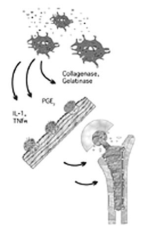
Introduction The recent surge of
interest and information in genetic research, sparked by the Human Genome
Project, have fostered the development of new techniques that have impacted
basic molecular biology and clinical medicine. Information gained from
these studies has enabled researchers to identify genes which are involved
in specific disorders and diseases. By identifying which genes are expressed,
researchers can better understand the biochemical pathways of disease.
This basic level of understanding of the disease process has enormous
potential for the treatment and prevention of disease. Furthermore, this
information provides tremendous opportunities in the field of gene-based
therapies, as highly specific, small molecules can be developed to target
genes involved in the biochemical process for specific illnesses. If research
indicates that a gene or group of genes is activated by the onset of a
particular pathology, a therapy can be developed that will interfere with
the biochemical pathway and prevent that activation, thus rendering the
disease asymptomatic.
A variety of molecular biology techniques have enabled us to delve into the human genome. Some of these techniques are limited in the amount of genes that can be simultaneously studied. Northern analysis and RNase protection assay (RPA) can be used to study upregulation of gene expression, but require relatively large amounts of sample RNA and are restricted to investigations of a few genes at each time. Reverse transcriptase- polymerase chain reaction (RT-PCR) requires small samples, but the results are also restricted in the number of genes that can be studied, and this technique is affected by contaminations. In contrast, transcriptional profil-ing using microarray technology permits examination of the effects of experimental manipulation on the expression pattern of vast numbers of genes and gene families that control cell responses.(1) Depending on the system, anywhere from 500 to 10,000 genes can be studied simultaneously in each experi-ment. In addition, microarrays require only a small amount of sample RNA (~2 g) through the use of a primer mixture, spe-cific for every gene on the array, as opposed to non-specific oligo dT priming used in other molecular techniques. The microarrays can use either fluorescent or radioactive labeling to visualize the genetic expression. The intensity of the expres-sion can be quantified using software, and the transcriptional profile can be used to illustrate the gene pathways effected by the disease process or experimental treatment. With this new technology, it is possible to study the temporal expression of genes as they are activated in the transcription process .(1,2) Technology is currently readily available to permit investigators to develop their own arrays or disease-specific arrays or chips.
Monocytes were harvested
from 400 ml peripheral blood using sequential Percoll gradients. (4) After
an overnight adher-ence depletion, the monocytes were cultured with submicron
UHMWPE (mean diameter 1.6 Ö m; 66% < 1 Ö m), TiAlV (mean diameter 1 Ö
m) or with no debris as controls .(4,7) Based on prior optimization studies,
the particle dose represented two times the surface area of cells, equivalent
to challenge with approximately 40 particles per cell. The cell culture
was inter-rupted at 30 min, 1 h, 4 h, 8 h, and 24 h, and the total RNA
extracted.
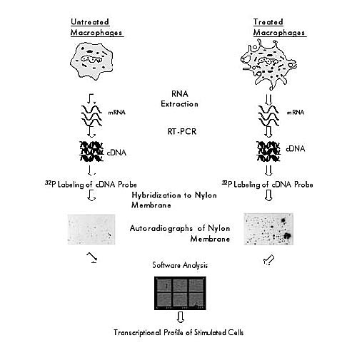
The probe mixture was reverse transcribed from the sample RNA using a mixture of primers specific for every gene on the array. The 32 P labeled probe mixture was hybridized to the arrays representing about 1,200 cDNAs and analyzed by autoradiography (Atlas Human Gene Array 1.2, Clontech, Palo Alto, CA). To determine the relative expression of a given gene, the autoradiographs were scanned and analyzed by array spe-cific software (AtlasImage 1.0, Clontech). A brief schematic of the procedure is provided in Figure 2.
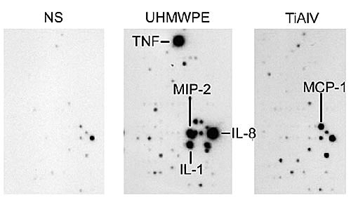
Results Both UHMWPE and TiAlV particle treatments produced marked increases in multiple pro-inflammatory cytokines including IL-1 , TNF-Í, and IL-8. Figure 3 illustrates hybridization patterns for IL-8, TNF-Í, and IL-1 after particle treatment. mRNA levels of many other genes of interest showed striking changes in steady-state levels, including message for several different chemokines and kinases involved in the regulation of cell responses. Most striking were the differences in the diversity and level of gene expression in UHMW-PE compared to TiAlV. Discussion These studies show the utility of the microarray technology for exploring the complex regulatory pathways and gene products involved in particle induced macrophage activation. This technology has the advantage of providing semi-quantitative screening of multiple classes of genes, including the gene regulation of cytokine expression, cell proliferation, and apoptosis. Furthermore, by comparing data from several different time periods, we have illustrated the temporal activation of these genes. Of particular interest was the finding that the gene expression was critically dependent upon the composition of the challenging particles. This approach provides a powerful technology for dissecting the molecular mechanisms by which wear debris regulates transcription of cytokine genes, related pro-inflammatory mediators, and contributes to aseptic loosen-ing of orthopaedic implants. By identifying which genes are involved in aseptic loosening and osteolysis, we can begin to approach joint arthroplasty from a different perspective. One possibility is to geno-stratify patients and develop drug thera-pies based on the genes patients exhibit and the response observed in vitro. If the activity of a gene or group of genes is successfully identified, therapies can be synthesized to target that gene and suppress its expression or target its product and inactivate it. Through such clinical studies, class ifications can be made as to which geno-stratified group would be more appropriate with which biomaterial (or its modifica-tion), thus increasing the success of the joint arthroplasty and reducing the need for future revision surgeries. Similar strate-gies may hold promise for other areas of orthopaedics such as fracture healing and tumor classification and treatment. In the near future, transcriptional profiling holds distinct opportunities to improve patient care and treatment in orthopaedics. Acknowledgments The authors would like to thank Jennifer Kenney for her technical assistance. The study was supported by NIH Grant DK 46773 (SRG) and the Department of Orthopaedic Surgery, Massachusetts General Hospital. |
||||||||
|
||||||||
| HOJ HOME | Chiefs Reports | Osgood Day | Scientific Articles | Alumni Association |
| References | |
| 1. | Iyer, V. R., M. B. Eisen, D. T. Ross, G. Schuler, T. Moore, J. C. F. Lee, J. M. Trent, L. M. Staudt, J. Hudson Jr., M. S. Boguski, D. Lashkari, D. Shalon, D. Botstein, and P. O. Brown. The transcriptional program in the response of human fibroblasts to serum. Science 283:83-87 (1999). |
| 2. | Schena, M., D. Shalon, R. W. Davis, and P. O. Brown. Quantitative monitoring of gene expression patterns with a complementary DNA microarray. Science. 270:467- 469(1995). |
| 3. | Shanbhag, A. S., D. R. Cho, K. Kas, J. H. Herndon, H. E. Rubash, and S. R. Goldring. The transcriptional response program of human monocyte activation by poly-ethylene. Trans. Orthop. Res. Soc. 46:52(2000). |
| 4. | Shanbhag, A. S., J. J. Jacobs, J. Black, J. O. Galante, and T. T. Glant. Human monocyte response to particulate biomaterials generated in vivo and in vitro. J. Orthop. Res. 13:792-801 (1995). |
| 5. | Shanbhag, A. S., C. T. Hasselman, and H. E. Rubash. Inhibition of wear debris mediated osteolysis in a canine total hip arthroplasty model. Clin. Orthop. 344:33-43 (1997). |
| 6. | Jiranek, W. A., M. Machado, M. J. Jasty, D. Jevsevar, H. J. Wolfe, S. R. Goldring, M. J. Goldberg, and W. H. Harris. 6. Jiranek, W. A., M. Machado, M. J. Jasty, D. Jevsevar, H. J. Wolfe, S. R. Goldring, M. J. Goldberg, and W. H. Harris. Production of cytokines around loose cemented acetabular components. Analysis with immunohistochemical techniques and in situ hybridization. J. Bone Joint Surg. 75-A: 863-879 (1993). |
| 7. | Shanbhag, A. S., C. T. Hasselman, and H. E. Rubash. Techniques for generating submicrometer ultra high molecular weight polyethylene. J. Orthop. Res. 14:1000- 1004 (1996). |
|
TOP OF PAGE | HOJ HOME |
| HOJ HOME | Chiefs Reports | Osgood Day | Scientific Articles | Alumni Association |