Investigating the Macrophage Response to Wear Debris Using High-throughput Protein Chips: Intense Inflammatory Reaction to Titanium-Alloy Particles
Adam M. Kaufman, Rajiv Sethi MD, Harry E. Rubash MD and Arun S. Shanbhag PhD, MBA
Massachusetts General Hospital
Introduction
Particle-induced bone resorption represents a
significant clinical problem in total joint arthroplasty,
accounting for the majority of long-term implant
failures (1;2). Submicron wear debris from both
the metallic and polymeric prosthetic components
is believed to stimulate resident macrophages to
release a variety of inflammatory cytokines. These
mediators recruit and activate inflammatory cells,
orchestrating the formation of a peri-implant granuloma.
This granuloma serves as a permanent source
of cytokines, ultimately causing osteoclastic bone
resorption and implant loosening.
In-vitro models using human macrophages remain the
gold standard for investigating the acute response to wear
debris (3;4). Researchers have classically relied on ELISA to
identify individual proteins such as TNF-a, IL-1 and IL-6 as the
primary drivers of osteolysis (5;6). This technique is simple,
but labor intensive limiting studies to a handful of cytokines.
Recent investigations using gene expression profiling demonstrate
that particle stimulation of macrophages is incredibly
complex and involves immediate expression of hundreds of
macrophage genes(7). In this study, we utilized emerging
high-throughput protein chips to characterize the short-term
response of human macrophages to clinically relevant wear
debris such as UHMWPE, TiAlV, CoCr and alumina particles.
Protein chips permitted the simultaneous and precise quantification
of 29 cytokines, chemokines and growth factors.
METHODS
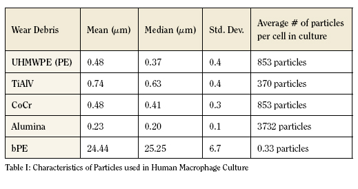
Particle Preparation
Fine UHMWPE, and TiAlV debris were prepared as previously
described (8;9). CoCr and Alumina particles were obtained
from commercial sources (Zimmer, Inc and Polysciences).
Particles were characterized using a JEOL 5910 scanning electron
microscope 10,000 – 15000 x magnification. Approximately
300 particles were measured for each species (Table I).
Cell Iso lation and Culture
Human monocytes were isolated from peripheral blood
donated by healthy volunteers (n=4) and purified by sequential
discontinuous Percoll gradients (10). 2 x 10⁶ cells/well were cultured
in macrophage serum-free media (M-SFM) supplemented
with antibiotics. After an overnight culture and washing to
remove non-adherent cells, adherent macrophages were challenged
with various particle species (see Table I) in 1 ml media
at concentrations representing 2x the macrophage surface area
(3). Controls included non-stimulated cells and lipopolysaccharide
(LPS) endotoxin served as a positive control. After a
24h culture, macrophage conditioned media were harvested,
aliquoted and stored at (-) 76OC.
High-throughput Protein Chips and Analysis
Monocyte supernatants were analyzed using the protein
profiling human Cytokine Biochip (Zyomyx, Hayward, CA).
Each chip contained five replicates and one internal control. 40
µl of sample was analyzed for each determination of 29 cytokines
in five replicates. Biochips were scanned on a fluorescent
scanner with a 532 nm laser and parameters adjusted to provide
the largest dynamic range with minimal feature saturation.
Protein concentrations were calculated based on a sixpoint
calibration curve performed in each chip. Each reported
value represents the average of five replicate measurements
from each assay. Data is presented as the ratio of particle treatment
over non-stimulated controls at time zero.
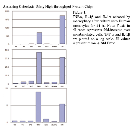
RESULTS
Data is presented and discussed in terms of fold-increase
over non-stimulated (NS) macrophages from the same donor
cultures (Figures 1 and 2). NS samples exhibited minimal cytokine
activity, suggesting that they were not activated during the
isolation procedure and thus validating the methodology. LPS
used as a positive control in these studies because it provides
the most robust stimulus to macrophages, attesting to their
viability and responsiveness in culture.
Nine of twenty-nine (9/29) cytokines included on the chip
were detected after the 24 h culture period. IL-1a, TNF-a and
IL-1ß stimulation was seen in select samples (Figure 1). TNF-
a release was stimulated by TiAlV (2000-fold), CoCr (22-fold),
bPE (22-fold) and alumina (5-fold). Similar trends were seen
in IL-1ß expression and release as TiAlV elicited a 30-70 fold
increase. CoCr and alumina particles elicited nominal increases
of 2-fold and 3.1-fold increases over controls, respectively. Only
TiAlV particles elicited IL-1a, almost 20-fold higher than control
macrophages. Submicron PE particles were surprisingly
benign, causing only an ~ 3-fold increase in IL-1ß production
with no significant IL-1a or TNF-a stimulation.
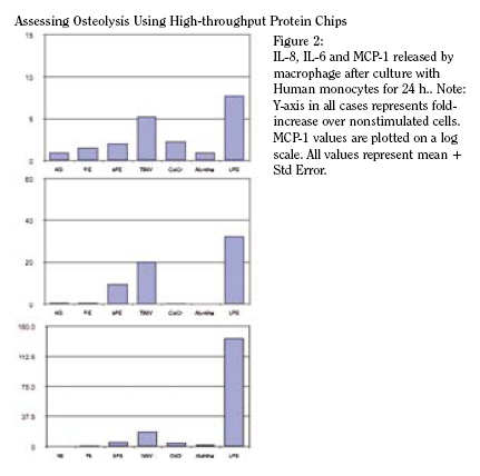
IL-8, IL-6, MCP-1 (Figure 2) were also detected. IL-6 was
stimulated only by TiAlV at levels ~ 20-fold higher than control
macrophages. TiAlV and bPE particles stimulated macrophages
to release nearly 5-fold and 2-fold higher level of IL-8. CoCr
and PE particles again elicited minimal increases relative to NS
cells. TiAlV also stimulated the highest levels of MCP-1 (18-fold
increase) compared to CoCr (5-fold), bPE (5-fold) and alumina
(3-fold) particles. GM-CSF and IL-10 were stimulated only by
TiAlV particles.
DISCUSSION
Wear debris-stimulated macrophages have long been
focused on as the primary driver of peri-implant loosening.
Our recent gene array studies portrayed a complex macrophage
response, as transcriptional alterations were made in hundreds
of genes immediately following interaction with wear debris (7).
The use of protein arrays in this study allowed us to simultaneously
and precisely quantify 29 protein products of these genes,
providing an accurate portrait of the acute response of human
macrophages to wear debris.
Of the 29 cytokines targeted, 9 were detected consistently
in the macrophage cultures. The absence of T-cell derived
mediators (IL-2, IL-3, IL-4, IL-4, IL-12p40, IL-15, IL-13,
SCD95, SCD23, IFN?) is not surprising and alludes to the purity
of isolated macrophages. IP-10 and MIG, are present in osteolytic
tissues (11), requires T-cell release of IFN?, not present in
our model here.
A surprising finding in this study was the intense inflammatory
response documented following macrophage challenge
with TiAlV particles. TiAlV was the only particle species to
cause a significant increase in GM-CSF, suggesting that the
acute exposure to macrophages alone is enough to generate
a systemic inflammatory response. TiAlV particles universally
elicited the highest levels of cytokines and the expression of
the classic osteolytic mediators TNF-a, IL-1a and IL-1ß, it
rivaled that of the positive control LPS. CoCr, alumina and
bPE wear caused inflammation of intermediate intensity. The
reaction to the cauliflower-shaped, 25 µm bPE confirms that
inflammation can occur independent of phagocytosis. The
relatively brisk reaction to TiAlV has been documented in the
literature. Comparing the macrophage response to CoCr and
TiAlV, Haynes et al reported that while CoCr particles released
basal levels of mediators, TiAlV particles elicited very high
levels of several mediators including PGE2, IL-1, TNF and IL-6
(12). In an earlier study using ELISA, Shanbhag et al reported
that human macrophages increase secretion of no more than
2-3-fold after TiAlV stimulation (10). Compared to this body of
literature, results from our current studies are consistent with
an unprecedented response to TiAlV wear debris.
There are several possible explanations for this increased
response. As we have recently demonstrated, TiAlV disks also
stimulate macrophages and elicit high levels of TNF-a, IL-6
and IL-1ß (13). This suggests the TiAlV surface chemistry and
texture are stimulatory, independent of phagocytosis (13). The
morphology of TiAlV debris is typically globular with some rodshaped
particles and flakes (14;15). In contrast, PE wear debris
are predominantly spherical with occasional fibrils interconnecting
debris aggregates (14).
TiAlV particles represent only 5% of wear released in vivo
while PE represents the majority, over 70-95% (14). As such,
engineering efforts to improve implants have focused on creating
wear resistant polymers rather than preventing metallic
debris. The results of this study suggest that PE debris is
relatively benign, eliciting cytokine levels only slightly higher
than controls. In contrast, the macrophage response to TiAlV
rivaled that of LPS and was generally more than 100 times
as stimulatory as PE. Metallic wear can also decrease healing
through metabolic alterations, osteoblastic inhibition (16),
chemical carcinogenesis (17), decreased mineralization (18)
and immunological interactions as hapten formation and
anti-chemotactic action (19). It is possible that these rare, but
stimulatory TiAlV particles could play a role equivalent to the
abundant but benign PE particles in peri-implant loosening.
While the emphasis on PE has resulted in developing wear
resistant UHMWPE, a similar effort needs to be expended to
reduce all sources of metal debris at the implant site.
Adam Kaufman is a third year medical student at Harvard Medical School.
Rajiv Sethi M.D. is a PGY-5 resident in the Harvard Combined Orthopaedic Residency.
Harry E. Rubash M.D. is Chief of the Orthopedic Department at Massachusetts General Hospital.
Arun S. Shanbhag Ph.D., MBA is Director of the Biomaterials Lab at Massachusetts General Hospital and Assistant Professor of Orthopedic Surgery at Harvard Medical School.
Address correspondence to:
Arun Shanbhag, Ph.D, MBA
GRJ 1115, 55 Fruit St.
Boston, MA 02114
shanbhag@helix.mgh.harvard.edu
References:
- Shanbhag AS, Hasselman CT, Jacobs JJ, Rubash HE. Biologic response to wear debris. In: Callaghan JJ, Rosenberg AG, Rubash HE, editors. The Adult Hip. Philadelphia, PA: Lippincott-Raven Publishers, 1998: 279-288.
- Ingham E, Fisher J. The role of macrophages in osteolysis of total joint replacement. Biomaterials 2005; 26(11):1271-1286.
- Shanbhag AS, Jacobs JJ, Black J, Galante JO, Glant TT. Macrophage/particle interactions: effect of size, composition and surface area. J Biomed Mater Res 1994; 28(1):81-90.
- Shanbhag AS, Macaulay W, Stefanovic-Racic M, Rubash HE. Nitric oxide release by macrophages in response to particulate wear debris. J Biomed Mater Res 1998; 41(3):497-503.
- Glant TT, Jacobs JJ, Molnar G, Shanbhag AS, Valyon M, Galante JO. Bone resorption activity of particulate-stimulated macrophages. J Bone Miner Res 1993; 8(9):1071- 1079.
- Schwarz EM, Lu AP, Goater JJ, Benz EB, Kollias G, Rosier RN, Puzas JE, O’Keefe RJ. Tumor necrosis factor-alpha/nuclear transcription factor-kappaB signaling in periprosthetic osteolysis. J Orthop Res 2000; 18(3):472-480.
- Garrigues GE, Cho DR, Rubash HE, Goldring SR, Herndon JH, Shanbhag AS. Gene expression clustering using self-organizing maps: analysis of the macrophage response to particulate biomaterials. Biomaterials 2005; 26(16):2933-2945.
- Shanbhag AS, Hasselman CT, Rubash HE. Technique for generating submicrometer ultra high molecular weight polyethylene particles. J Orthop Res 1996; 14(6):1000- 1004.
- Yang IH, Kim SY, Rubash HE, Shanbhag AS. Fabrication of submicron titanium-alloy particles for biological response studies. J Biomed Mater Res 1999; 48(3):220-223.
- Shanbhag AS, Jacobs JJ, Black J, Galante JO, Glant TT. Human monocyte response to particulate biomaterials generated in vivo and in vitro. J Orthop Res 1995; 13(5):792-801.
- Shanbhag AS, Kaufman AM, Agarwal S, Hayata K, Decker J, Kawashima M, Freiberg A, Grills G, Rubash HE. Critical Insights into osteolysis using protein microarrays: The importance of IL-6 and T-cell Activation. Trans Orthop Res Soc 2005; 30:148.
- Haynes DR, Rogers SD, Hay S, Pearcy MJ, Howie DW. The differences in toxicity and release of bone-resorbing mediators induced by titanium and cobalt-chromiumalloy wear particles. J Bone Joint Surg Am 1993; 75(6):825-834.
- Sethi RK, Neavyn MJ, Rubash HE, Shanbhag AS. Macrophage response to cross-linked and conventional UHMWPE. Biomaterials 2003; 24(15):2561-2573.
- Shanbhag AS, Jacobs JJ, Glant TT, Gilbert JL, Black J, Galante JO. Composition and morphology of wear debris in failed uncemented total hip replacement. J Bone Joint Surg Br 1994; 76(1):60-67.
- Maloney WJ, Smith RL, Schmalzried TP, Chiba J, Huene D, Rubash H. Isolation and characterization of wear particles generated in patients who have had failure of a hip arthroplasty without cement. J Bone Joint Surg Am 1995; 77(9):1301-1310.
- Vermes C, Roebuck KA, Chandrasekaran R, Dobai JG, Jacobs JJ, Glant TT. Particulate wear debris activates protein tyrosine kinases and nuclear factor kappaB, which down-regulates type I collagen synthesis in human osteoblasts. J Bone Miner Res 2000; 15(9):1756-1765.
- Gillespie WJ, Frampton CMA, Henderson RJ, Ryan PM. The incidence of cancer following total hip replacement. J Bone Joint Surg 1988; 70-B:539-542.
- Blumenthal NC, Cosma V. Inhibition of apatite formation by titanium and vanadium ions. J Appl Biomat 1989; 23:13-22.
- Merritt K, Brown SA. Hypersensitivity to metallic biomaterials. In: Williams DF, editor. Systemic Aspects of Biocompatibility, vol. II. Boca Raton, FL: CRC Press, 1981: 33-48.
|