|
|
|
| Click here to visit our web site |
| INTRODUCTION
Despite advancements in spinal instrumentation and bone grafting techniques, clinical results of spinal fusions are often negatively influenced by the development of pseudoarthrosis. Electrical stimulation is now a popular technique to enhance bone formation in spinal fusions. The current methods used to enhance spinal fusion include direct current electrical stimulation, capacitive coupling, combined magnetic fields, and pulsed electromagnetic fields. (1-3) HISTORY OF ELECTRICAL STIMULATION
The modern theories of electrical stimulation are based on the work of Yasuda. In 1953, Yasuda and his colleagues determined that areas of bone in compression were electronegative, while areas in tension were electropositive. They proposed that electrical energy imparted to a bone would initiate callus formation. Using direct current, they showed in a rabbit model the formation of a ridge of callus along the cathode, and confirmed their theory. (7,8) Yasuda’s observations were independently confirmed by multiple investigators. (9-11) Friedenberg and Brighton further elucidated that in areas of bone undergoing active growth or repair, the potential is electronegative in relation to areas of resting bone. (12) This awareness that bone responds to electrical stimulation encouraged further investigations into the effects of electrical stimulation on bone formation and growth. VARIOUS METHODS OF ELECTRICAL STIMULATION
DIRECT CURRENT ELECTRICAL STIMULATION
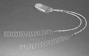
Direct current stimulation involves the surgical implantation of electrodes with a battery (Figure 1). The cathode is placed in direct contact with the proposed site of fusion. The cathodes’ effective stimulation distance is 5 to 8 mm. By coiling the cathode, one can enhance the surface area and stimulation bed. The batteries deliver a constant, direct current for 6 to 9 months. Direct current obviates the need for patient compliance. However, there are some disadvantages to intracorporal battery placement. First, battery placement takes approximately 10-15 minutes, thereby lengthening the time that the patient is under general anesthesia. Secondly, the manufacturer suggests that the battery be removed in six months. Although the battery is placed in the subcutaneous space and can be removed under local anesthesia, this does entail a second surgical procedure for the patient. Finally, there is a risk of seeding the battery and the leads from a systemic infection. CAPACITIVELY COUPLED ELECTRICAL STIMULATION
Capacitive coupling uses a pair of external
electrodes PULSED ELECTROMAGNETIC FIELDS
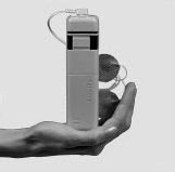
The use of pulsed electromagnetic fields requires neither additional intraoperative time nor a second surgical procedure for battery removal. The system consists of a noninvasive external coil that delivers electromagnetic energy when driven by an electric current. This method does require good patient compliance. The external units require six to eight hours of use each day in order to be clinically effective. No adverse systemic effects have been reported with the use of these external devices. EXPERIMENTAL STUDIES In cell cultures, direct current is capable of inducing cell division and recruitment of osteogenic cells. In vivo, osteogenesis is thought to be due to a reaction at the electrodetissue interface. Water undergoes hydrolysis, reducing oxygen tension, which in turn produces hydroxyl ions. This reaction increases the local tissue pH. An alkaline and low PO2 environment is thought to be optimal for bone formation. Recent work has revealed that specific signal transduction occurs in electrically stimulated bone cells. Brighton et al. evaluated the use of capacitive coupling, inductive coupling, or combined electromagnetic fields on cultured bone cells. All three techniques led to an increase in cytosolic Ca2+ and in activated cytoskeletal calmodulin. This increase in activated calmodulin has been shown to increase cell proliferation. (18) An understanding of the signal transduction and metabolic pathways induced by electrical stimulation will improve the ability of researchers and clinicians to utilize electricity to enhance spinal fusion. ANIMAL STUDIES OF DIRECT CURRENT ELECTRICAL
STIMULATION Kahanovitz and Arnoczky performed a well-controlled study on canine lumbar spinal fusions in 1990. They performed bilateral facet fusions at L1-L2 and L4-L5 in 12 dogs. Six animals received direct current electrical stimulation, and six received inactive devices. At 12 weeks, the animals that had received the stimulation demonstrated complete arthrodesis. (20) In 1999, Bozic et al. published the results of a rabbit fusion model, using coralline hydroxyapatite as a bone graft substitute in combination with direct current electrical stimulation. There were four experimental groups: one had autologous bone alone; the second had coralline hydroxyapatite with a proximal tibia aspirate; the third had coralline hydroxyapatite with an iliac crest aspirate and 40 microamps of direct current electrical stimulation; and the fourth had coralline hydroxyapatite with an iliac crest aspirate and 100 microamps of direct current electrical stimulation. The animals were sacrificed at eight weeks and underwent radiographic, histologic and mechanical testing. The results demonstrated a dose-dependent response to the direct current stimulation. The 100 microamp device significantly enhanced fusion success. The combination of coralline hydroxyapatite and the 100 microamp device achieved better fusion rates than that of autologous bone graft alone. (21) Toth et al, in 1999, used varying doses of direct current electrical stimulation on an interbody fusion model in sheep. This study involved a single level discectomy and fusion with a titanium cage. This study involved three groups of animals: one group underwent cage application with an inactive stimulator; the second group had 30 microamps; and the third group had 100 microamps of stimulation. The authors discovered an enhanced fusion rate and biomechanically stiffer fusion masses in the 100 microamp group. (22) Another recent study by Kahanovitz et al. evaluated the effect of varied current densities on spinal fusion. Using his original canine fusion model, Kahanovitz demonstrated a doseresponse of fusion mass scores to increasing current density. The highest current density (10 microamps/cm) showed a statistically higher fusion score than the lowest current density (0.83 microamps/cm) at six and nine weeks. No differences were noted at 12 weeks as all groups showed complete fusion. This study suggests that the speed of fusion may be improved by increasing electrical stimulation current density. (23) ANIMAL STUDIES OF PULSED ELECTROMAGNETIC FIELDS
In 1994, Guizzardi et al. reported an enhancement of bone formation with PEMF stimulation in a rat model at four and eight weeks. (25) Glazer et al. studied the effect of PEMFs on a rabbit spinal fusion model. The authors performed a wellcontrolled study, comparing autograft alone or with PEMF. Over the course of six weeks, for four hours each day, the animals were placed in cages designed to create a uniform field over the fusion site. The analysis included biomechanic, histologic, and radiographic evaluation of the fusion masses. The study demonstrated that the stiffness of the fusion mass treated with PEMF was 37% greater than that of controls. Furthermore, an enhanced fusion rate was seen with PEMF, but it was not statistically significant. (26) Clinical Studies The results were statistically significant, with a successful fusion rate of 91% in the stimulated group, versus 81% in the control group. Kane’s second trial was a randomized prospective controlled trial of electrical stimulation in “difficult” spinal fusion patients. He defined the “difficult” patients as those with at least one of the following: 1) one or more previous failed spinal fusions, 2) Grade II or worse spondylolisthesis, 3) extensive bone grafting necessary for a multiple-level fusion, or 4) other high-risk factors for failure of fusion, including gross obesity. Again the results were statistically significant: there was a successful fusion rate of 81% for the stimulated group versus 54% for the controls. Finally, the third trial analyzed the results of an uncontrolled group of “difficult” patients who had undergone posterior spinal fusion supplemented with direct current electrical stimulator implantation. The fusion rate was 93%. (29) In 1994, Meril reported a 93% rate of successful fusion in patients who had undergone either anterior or posterior lumbar interbody fusion with electrical stimulation. The control patients achieved only a 75% fusion rate. In Meril’s study, four of the 122 patients who had received implantable stimulators required removal of the device during the course of treatment secondary to patient discomfort. (30) Pettine performed a retrospective study of DC current stimulation in 1995. He found a higher fusion rate in those patients receiving electrical stimulation (89% versus 84% overall). In “high-risk” patients, there was a fusion rate of 96% in the stimulated group versus 80% in controls. In 1996, Rogozinski and Rogozinski published a study which analyzed the efficacy of electrical bone growth stimulation in spinal fusion surgery using pedicle screw fixation. The patients all underwent posterolateral fusion with autologous bone graft and fixation using a pedicle screw and rod system. The patients had been matched for diagnostic criteria, surgical approach, fixation technique, and rehabilitative regimen. Fusion surgery was performed by the same two surgeons for all patients. The average follow-up was 20. 5 months. The group that received electrical stimulation achieved a 96% successful fusion rate versus an 85% fusion rate for control patients. The higher absolute rates of successful fusion achieved in the Rogozinski study when compared with Kane’s study implies increased fusion rates when instrumentation is used. (31) Kucharzyk reported the results of a controlled prospective outcome study in a high-risk fusion population. A total of 130 patients underwent posterolateral spinal fusion with autologous graft and pedicle screw fixation instrumentation. The patients were divided into two 65-patient groups, with one group receiving an implantable electrical stimulator and the other group acting as a control. The average overall follow-up period was 3.8 years. Patient follow-up examinations included radiographic evaluation of the lumbar spine for assessment of fusion. The radiographic criteria for successful fusion were consolidation of bone graft, observation of bridging trabeculae, absence of pseudarthrosis lines, absence of correlating pain, and absence of instrumentation failure. In addition, patients were evaluated for clinical outcome using the modified Smiley- Webster surgical rating scale; only ratings of excellent or good were accepted as successful. Kucharzyk reported a successful fusion rate of 95. 6% in stimulated patients versus 87% in the control group. Clinical outcome evaluation demonstrated a 95% success rate in the stimulated group versus 79% in the control group. (32) In 1996, Tejano et al. reviewed a series of patients who had undergone a posterolateral intertransverse-process or facet fusions with autogenous iliac crest bone graft in conjunction with an EBI electrical stimulator DC current device. The overall fusion success rate was 91.5%. A lower fusion rate was seen in those patients undergoing pseudarthrosis repair (80%). (33) CLINICAL STUDIES OF PULSED ELECTROMAGNETIC FIELDS
Lee described a series of patients undergoing pseudarthrosis repair using PEMF postoperatively, in 1989. He reported a 67% success rate. (35) Soon after, in 1990, Mooney analyzed postoperative PEMF treatment in patients who had undergone either anterior or posterior lumbar interbody fusions. Radiographic fusion success was defined as 50% incorporation of the fusion mass. Patients who were compliant with the use of the PEMF device had a successful fusion rate of 92.2%. Those who were noncompliant with the use of the PEMF device and those with the placebo device, had fusion rates of 64. 9% and 67.9%, respectively. Interestingly, internal fixation, smoking, as well as type of bone graft used (allograft versus autogenous iliac crest) had no effect on the fusion rate. (36) Linovitz presented the results of a double-blind, randomized, placebo-controlled study on PEMF in 2000. Patients underwent single or two-level lumbar fusions without instrumentation. Patients were randomized to receive either a placebo or an active PEMF unit postoperatively. Only one half-hour per day of PEMF stimulus was used for nine months. The results demonstrated a higher fusion rate of 64% in the treated group versus 43% in the placebo group. (37) CLINICAL STUDIES OF CAPACITIVE COUPLING
FUTURE STUDIES
Paul Andrew Glazer, MD is a Clinical Instructor in Orthopaedic Surgery at Harvard Medical School. Liane Clamen Glazer, MD is a Resident, Department of Ophthalmology, Massachusetts Eye and Ear Infirmary, Harvard Medical School. Address correspondence to: |
|
Print Manuscript • View References • Download PDF version • Close window |