|
|
|
| Click here to visit our web site |
|
Posterior-substituting (PS) total knee arthroplasty (TKA) was introduced to improve knee flexion by inducing tibiofemoral rollback (posterior femoral translation), and to prevent posterior subluxation of the tibia. (7,11) The femoral cam is designed to engage the tibial spine of the prosthesis during knee flexion, in order to prevent excessive posterior tibial translation and posterior tibial impingement with flexion, thus increasing the maximum flexion of the TKA. However, clinical studies have demonstrated that maximum flexion after PS TKA is usually less than 120°. (3) The few in vivo biomechanical studies that have investigated the mechanisms of this cam-spine system interaction have yielded inconsistent results on the ability of PS TKA to restore normal knee kinematics. Most have reported reduced tibiofemoral rollback when compared to normal knees. (1,2,5,12) One drawback of these studies is that they were not able to compare motion of the same knee before and after TKA. It is therefore difficult to objectively evaluate the ability of TKA to restore normal knee kinematics. Moreover, limited quantitative data have been reported on when and how the cam-spine mechanism is effective during flexion-extension of the knee. This information would be invaluable as a basis for further improvement of TKA design, in order to achieve high flexion of the knee (up to 160°) after TKA. In this study, we tested a series of human cadaveric knee specimens using the robotic testing system, to investigate the kinematic responses of the knee before and after a posterior substituting total knee replacement (NexGen, Zimmer, Inc. ) under simulated muscle loading conditions. Using the principle of superposition, we also determined the cam-spine contact forces as a function of knee flexion and loading condition. Knee kinematics were defined in terms of translation of the lateral and medial femoral condyles on the tibial plateau, from which both anterior-posterior femoral translation and internal-external tibial rotation could be derived. Quadriceps (400N) and hamstring (semimembranosus, semitendinosus and biceps femoris, 200N total) muscle forces were simulated using a pulley system and weights attached to the appropriate muscle tendons.
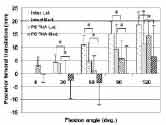
We found that, in the unloaded condition, posterior translation of the lateral and medial femoral condyles after TKA was significantly reduced compared to that of the native knee at all flexion angles except at full extension (Figure 4). At 120° of flexion, posterior translation of the lateral femoral condyle was reduced by 23%, and posterior translation of the medial condyle was reduced by 40%. Similar trends were observed under simulated muscle loads, as posterior translation of both medial and lateral femoral condyles were reduced when compared to that of the intact knee. Comparison of the kinematics of unloaded and muscle loading conditions at higher flexion angles (90° and 120°) revealed that medial and lateral condyle translations did not differ significantly with the addition of simulated muscle loads. Contact forces between the femoral cam and tibial spine ranged from 20N to 90N (Figure 5). Forces were lowest without simulated muscle loads during the passive path. Under simulated muscle loads, increasing contact forces were seen with increasing knee flexion, with highest contact forces observed under combined quadriceps and hamstring loading at 120° and under isolated hamstrings loading at 90°.
Our findings are consistent with reported in vivo knee kinematics after TKA during various activities. (2,12,13) Reduced posterior femoral translation of PS TKA during step-up and gait has been reported in in vivo studies,(9,13) which corresponds with our findings of reduced posterior femoral translation after PS TKA under simulated muscle loads. The cam-spine contact forces measured at low flexion angles were approximately 20N. Fluoroscopy performed during experiments revealed that the cam-spine mechanism was not engaged at these low flexion angles, suggesting that the recorded forces at these angles were not due to direct contact of the posterior surface of the spine with the femoral cam. We believe that this residual force arose from contact of the sides of the spine with the femoral component. Engagement of the cam-spine mechanism with knee flexion beyond 70° partially restored posterior femoral translation. Reduced posterior translation of the femoral condyles after PS TKA may limit knee flexion by early impingement of the femoral shaft with the posterior edge of the tibial component. This loss of posterior femoral rollback may account for clinical observations of the inability to achieve knee flexion beyond 120° after PS TKA. According to our results, reduced femoral translation at high flexion angles after PS TKA is independent of muscle loads. Therefore, optimized prosthetic design and/or surgical technique may be important in determining proper timing of the cam-spine engagement to increase knee flexion. The position of the polyethylene spine on the tibial plateau is an important factor influencing the timing of the cam-spine engagement. Using a computer model, Delp et al. reported a posteriorly positioned tibial spine component will cause early cam-spine engagement, thus increasing the extent of posterior femoral translation. (4) Similarly, different geometrical designs of TKA components as well as variation in surgical techniques could all influence cam-spine behavior and potential knee flexion after PS TKA. In summary, we investigated the mechanism of the camspine system using a single design posterior-substituting total knee arthroplasty. We found that the cam-spine system was partially effective at restoring tibiofemoral rollback at increasing flexion angles, but did not completely restore posterior rollback to that of the intact knee. This lack of posterior rollback may limit the ability of PS TKA to restore flexion beyond 120° due to early tibiofemoral impingement. We hope that our work will help to develop a rationale for further improvement of surgical technique and prosthetic designs, with the ultimate goal of restoring normal knee function over the entire range of flexion after total knee arthroplasty. ACKNOWLEDGEMENTS This study was supported by a research grant from Zimmer, Inc. and a National Science Foundation (NSF) fellowship.
|
|
Print Manuscript • View References • Download PDF version • Close window |